Ligand-switchable nanoparticles resembling viral surface for sequential drug delivery and improved oral insulin therapy
T. Yang, A. Wang, et al.
Explore these studies to deepen your understanding
Adjacent work that informs or extends this paper's methodology and findings.
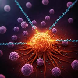
Nanoparticles and convergence of artificial intelligence for targeted drug delivery for cancer therapy: Current progress and challenges
R. P. Singh, A. Natarajan, et al.
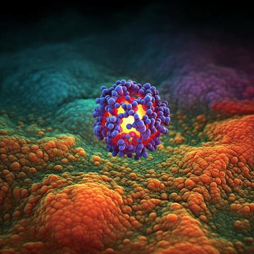
Nanoparticles exhibiting virus-mimic surface topology for enhanced oral delivery
Z. Sang, L. Xu, et al.
Highly porous and injectable hydrogels derived from cartilage acellularized matrix exhibit reduction and NIR light dual-responsive drug release properties for application in antitumor therapy
M. Gulfam, S. Jo, et al.

Coacervation in polyzwitterion-polyelectrolyte systems and their potential applications for gastrointestinal drug delivery platforms
K. O. Margossian, M. U. Brown, et al.


