BiologyNature Communications
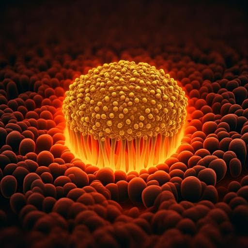
Life at high temperature observed *in vitro* upon laser heating of gold nanoparticles
C. Molinaro, M. Bénéfice, et al.
Discover how laser-assisted high-temperature microscopy (LA-HTM) is revolutionizing the study of thermophiles, those remarkable microorganisms thriving in extreme heat. Researchers Céline Molinaro, Maëlle Bénéfice, Aurore Gorlas, Violette Da Cunha, Hadrien M. L. Robert, Ryan Catchpole, Laurent Gallais, Patrick Forterre, and Guillaume Baffou showcase this innovative method that leverages gold nanoparticles to reveal the unique behaviors of *Geobacillus stearothermophilus* and *Sulfolobus shibatae* at elevated temperatures.
Related Publications
Explore these studies to deepen your understanding
Adjacent work that informs or extends this paper's methodology and findings.
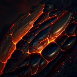
Earth Sciences
In situ observation of glass-like fragmentation of high-temperature silicate melts generating fine ashes
A. Namiki, S. Okumura, et al.
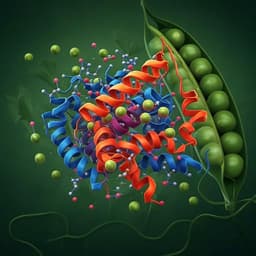
Food Science and Technology
Comparative effects of high pressure processing and heat treatment on in vitro digestibility of pea protein and starch
A. E. Hall and C. I. Moraru
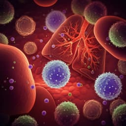
Medicine and Health
High Prevalence of Respiratory Co-Infections and Risk Factors in COVID-19 Patients at Hospital Admission During an Epidemic Peak in China
X. Zhu, F. Tian, et al.
Chemistry
Stabilization of gamma sulfur at room temperature to enable the use of carbonate electrolyte in Li-S batteries
R. Pai, A. Singh, et al.


