L-valine is a powerful stimulator of GLP-1 secretion in rodents and stimulates secretion through ATP-sensitive potassium channels and voltage-gated calcium channels
I. M. Modvig, M. M. Smits, et al.
Explore these studies to deepen your understanding
Adjacent work that informs or extends this paper's methodology and findings.
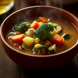
Validation of preferred salt concentration in soup based on a randomized blinded experiment in multiple regions in Japan—influence of umami (L-glutamate) on saltiness and palatability of low-salt solutions
H. Hayabuchi, R. Morita, et al.

Is Lennie a monster? A reconsideration of Steinbeck's Of Mice and Men in a 21st century inclusive classroom context
C. Lawrence

Diet, gym, supplements, or maybe it is all in your mind? A systematic review and meta-analysis of studies on placebo and nocebo effects in weight loss in adults
Ł. Kryst, P. Bąbel, et al.

Evaluating the effectiveness of the Kidogo model in empowering women and strengthening their capacities to engage in paid labor opportunities through the provision of quality childcare: a study protocol for an exploratory study in Nakuru County, Kenya
K. Okelo, M. Nampijja, et al.


