
Biology
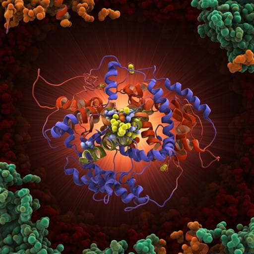
Key role of quinone in the mechanism of respiratory complex I
J. Gutiérrez-fernández, K. Kaszuba, et al.
Discover groundbreaking insights into respiratory complex I in this fascinating study by Javier Gutiérrez-Fernández and colleagues. The research reveals how quinone binding, rather than NADH, triggers significant structural changes that are crucial for understanding the energy coupling mechanism in this vital complex.
~3 min • Beginner • English
Introduction
The study addresses how NADH:ubiquinone oxidoreductase (complex I) couples electron transfer in the peripheral arm to long-range proton pumping in the membrane domain. Complex I is a large L-shaped enzyme with a redox chain from FMN through seven Fe–S clusters to the quinone (Q) site at the arm interface, and three antiporter-like subunits forming proton channels along a central hydrophilic axis. Although models have proposed conformational and/or electrostatic coupling (e.g., electrostatic spring), direct atomic-resolution evidence for conformational pathways linking the Q site to proton pumps has been lacking. The purpose is to determine structures of T. thermophilus complex I in multiple ligand-bound and native states to identify conformational changes triggered by substrates/inhibitors and to map how these changes propagate toward proton channels, clarifying the coupling mechanism and the role of quinone.
Literature Review
Prior structural work provided high-resolution bacterial and mitochondrial complex I structures, revealing the L-shaped architecture, Q-binding cavity near cluster N2, and antiporter-like subunits forming putative proton channels. Proposals for coupling include conformational propagation along a central hydrophilic axis and the electrostatic spring model where sequential Lys-Glu ion pair disruptions mediate pumping. Molecular dynamics and cryo-EM studies in mammals showed open/closed or active/deactive states with major loop rearrangements and TM3 rotation in subunit Nqo10/ND1. Quinone positions at intermediate and deep sites have been reported in other species. However, atomic-resolution comparisons of distinct redox/ligand states in a single system to define a conformational pathway from the Q site to proton channels remained incomplete.
Methodology
- Structural determination: Five X-ray crystal structures of T. thermophilus complex I were solved: native intact (CXIINT, updated PDB 6Y11), NADH-bound (CXINADH), and with Q-site ligands: decylubiquinone (CXIDQ), piericidin A (CXIPIE), aureothin (CXIAUT), pyridaben (CXIPYR). Crystals in space group P2₁ were refined at 3.1–3.6 Å with anisotropic truncation due to weak b-axis diffraction. Ligands were modelled where clear Fo–Fc and 2Fo–Fc density supported binding. A previously weak loop (Nqo6 55–70) was rebuilt using merged native datasets.
- Cryo-EM: Single-particle cryo-EM of complex I incubated with NADH or NAD+ prior to freezing yielded major and minor conformers regardless of nucleotide. Major classes were ~60% of particles at ~4.2–4.3 Å; minor ~40% at ~5.5–6.1 Å. Model building used MDFF and Rosetta-guided optimization with subsequent global optimization.
- Biochemistry: Enzyme activity with DQ was ~40 µmol NADH min⁻¹ mg⁻¹ and was fully inhibited by rotenone or piericidin A; IC50 values for inhibitors were measured (Supplementary). Crystallization with ligands employed co-crystallization and/or soaking under specified conditions; reducing conditions for NADH soaks used anaerobic glove box with sodium dithionite.
- Computational analyses: Normal mode analysis (NMA) using ANM/HCA elastic network models identified low-frequency modes and overlap analysis (OA) quantified contributions to experimental transitions; deformation analysis mapped hinges. Coarse-grained MD (Martini 2.2 with ElNeDyn restraints) simulated states with native-length ubiquinone in different redox/protonation forms (UQ, UQ²⁻, UQH2, and a state with negative charges on H38/Y87 donors), embedded in DOPC; two 2-µs replicates per state. Correlation network analysis of NMA and CG-MD trajectories identified allosteric communities and couplings. Water networks were modelled by assigning protonation states with PROPKA and placing waters with Dowser to evaluate Grotthuss-competent pathways.
Key Findings
- NADH binding alone does not induce global conformational changes: CXINADH is highly similar to CXIINT (global RMSD ~0.56 Å vs INT); after prolonged NADH soak, local changes occur—NADH shifts out of its canonical pocket, FMN density weakens, and key H38 of Nqo4 flips outward, potentially aiding quinol ejection.
- Cryo-EM reveals two conformers without Q-site ligand: major (~60%) and minor (~40%) states independent of NAD+/NADH presence; membrane domains align well while peripheral arm (PA) differs by hinge-bending rotation. CXIMJ resembles CXIINT; CXIMN resembles ligand-induced states. Global RMSDs vs INT: MJ ~2.00 Å, MN ~2.88 Å (membrane-domain RMSDs ~1.16–1.33 Å).
- Quinone-like ligands drive a conserved conformational response: All Q-site ligands bind at the deep site (~12 Å from N2) with a conserved H-bond from the quinone ketone to essential Y87. DQ causes the largest changes among ligands. Binding “tightens” the Q cavity; CASTp shows cavity surface area decreases 1703→1297 Ų and volume 997→812 ų upon DQ binding.
- Propagation pathway from Q site to E-channel: Ligand binding repositions Nqo4 β1–β2 loop (H38) ~1.5 Å inward and shifts Nqo8 TM5–6 loop (Q226/E227) ~3 Å into the cavity; Nqo8 TM helices tilt, transmitting motion to Nqo10 TM3 (Y59 region) which rotates ~30° (cytoplasmic half). These changes extend into Nqo11 (E32 moves ~1.2 Å from E67) and narrow the Nqo11–Nqo14 interface, potentially facilitating inter-subunit proton transfer. Changes beyond the E-channel into antiporter-like subunits were not observed under these non-turnover conditions.
- Water pathway reconfiguration: Only in CXIDQ, a single water is placed between E32 and E67 of Nqo11, forming a unique continuous path from the E-channel toward the first antiporter (Nqo14). This is enabled by Y59^10 rotation and subsequent E32^11 rotation, creating space for the water.
- Normal mode analysis: Two lowest-frequency modes observed experimentally: mode 7 (PA–MD bending) and mode 8 (PA rotation). OA shows transitions to CXIDQ involve large contributions from rotational mode 8; transitions among unliganded states involve primarily bending (mode 7). Hinges include the long Nqo7 loop at the PA–MD junction, the key Nqo4 β-sheet, and the C-terminus of Nqo12 traverse helix HL.
- Allosteric coupling networks: Community analysis confirms coupling between the Q-site region (Nqo4, and additionally Nqo6 in CXIDQ) and the E-channel node (Nqo7/10/11), strongest in CXIDQ among experimental structures.
- CG-MD supports charge-driven coupling: The strongest dynamic coupling between Nqo4 and the E-channel occurs in the modeled CXIUQH2_H38−/Y87− state (negative charges on proton-donating residues after Q reduction/protonation), stronger than in UQ or UQ²⁻ states. Detailed path analysis shows a unique hinge-region path via the Nqo4 β-sheet only in this charged-donor state, consistent with experimentally observed rotations.
- Comparative context: In T. thermophilus, TM3^10 rotates ~30°, smaller than ~180° reported for mammalian open/closed transitions, but the directionality and pattern are conserved across species.
Overall, quinone binding and subsequent chemistry, rather than Fe–S chain reduction alone, are the primary triggers of conformational changes that couple the Q-site to the E-channel, representing early steps in proton translocation.
Discussion
The findings directly address how energy from quinone reduction is coupled to proton pumping. Structural comparisons establish that binding of Q-site ligands induces a defined sequence of local rearrangements at the Q site that propagate through Nqo8 into the E-channel, accompanied by global PA bending/rotation. NADH-induced reduction without quinone does not produce these global changes, highlighting the central role of quinone. NMA and network analyses link these large-scale motions to localized changes in E-channel residues and hydrophobic surroundings. CG-MD indicates that charge transfer to the proton-donating residues (H38/Y87) upon quinone chemistry maximally strengthens allosteric coupling to the E-channel, suggesting that electrostatic effects of transiently charged donors synergize with conformational changes. The observed water-network reconfiguration (unique E32^11–H2O–E67^11 bridge in CXIDQ) provides a plausible structural basis for initiating proton transfer toward antiporter subunits. Together, the data support a mechanism wherein quinone binding tightens the Q cavity and rotates/bends the complex toward a minor-like state, priming the E-channel; subsequent quinone reduction/protonation imposes an electrostatically driven signal that advances switching of proton channels and proton ejection, likely completing under membrane and turnover conditions.
Conclusion
This study provides multi-state X-ray and cryo-EM structures of T. thermophilus complex I, revealing that quinone binding—rather than NADH-induced reduction—triggers conserved conformational changes that propagate from the Q site to the E-channel, accompanied by global PA motions. Computational analyses show that the strongest coupling to the E-channel arises when negative charge resides transiently on H38/Y87 during quinone chemistry, highlighting a central mechanistic role for quinone binding and protonation events. The work proposes a coherent coupling model with two low-energy global states (major-like and minor-like) connected by Q-driven transitions. Future work should capture high-resolution turnover states in membranes, quantify electrostatic versus conformational contributions within antiporter-like subunits, delineate periplasmic exit pathways, and assess effects of native long-tailed quinones and physiological gradients on the extent of conformational propagation.
Limitations
- Structural changes beyond the E-channel into antiporter-like subunits were not observed under the non-turnover crystallographic conditions; a full catalytic cycle, membrane environment, and pH gradient may be required.
- Cryo-EM minor states were at limited resolution (~5.5–6.1 Å), precluding detailed side-chain assignments (e.g., β1–β2 loop conformation) and water placement.
- NADH-soak crystals likely achieved full reduction only after long incubations, introducing potential heterogeneity and limiting temporal resolution of redox-dependent changes.
- Water-network analyses are based on modeled waters (Dowser) and predicted protonation states (PROPKA), not experimentally resolved waters at all sites.
- CG-MD uses coarse-grained force fields and DOPC bilayers which may not fully capture atomistic details or native membrane composition (menaquinone-8 in vivo).
- Allosteric network inferences rely on elastic-network/NMA and CG-MD correlations and do not directly measure proton flux or dynamics under turnover.
Related Publications
Explore these studies to deepen your understanding of the subject.


