
Medicine and HealthNature Metabolism
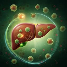
Itaconic acid underpins hepatocyte lipid metabolism in non-alcoholic fatty liver disease in male mice
J. M. Weiss, E. M. Palmieri, et al.
Discover the groundbreaking research by Jonathan M. Weiss and colleagues, which unveils the role of itaconic acid in mitigating lipid accumulation in liver diseases. This study investigates how macrophage-derived itaconate can potentially reverse adverse metabolic conditions in non-alcoholic fatty liver disease.
Related Publications
Explore these studies to deepen your understanding
Adjacent work that informs or extends this paper's methodology and findings.
Medicine and Health
Alantolactone attenuates high-fat diet-induced inflammation and oxidative stress in non-alcoholic fatty liver disease
J. Wang, Y. Jiang, et al.
Medicine and Health
Potential role of inflammation in relation to dietary sodium and β-carotene with non-alcoholic fatty liver disease: a mediation analysis
Y. Chen, M. Wu, et al.
Medicine and Health
Perilipin 5 regulates hepatic stellate cell activation and high-fat diet-induced non-alcoholic fatty liver disease
X. Yin, L. Dong, et al.
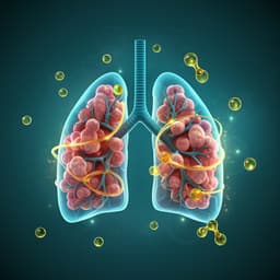
Medicine and Health
Omega-3 polyunsaturated fatty acids ameliorate PM2.5 exposure induced lung injury in mice through remodeling the gut microbiota and modulating the lung metabolism
J. Li, Y. Chen, et al.


