
Medicine and HealthCommunications Biology
Invadopodia enable cooperative invasion and metastasis of breast cancer cells
L. Perrin, E. Belova, et al.
This study reveals how different cancer clones work together to invade and metastasize, showcasing the vital role of invadopodia in leader cells. Conducted by Louisiane Perrin, Elizaveta Belova, Battuya Bayarmagnai, Erkan Tüzel, and Bojana Gligorijevic, the research highlights the intriguing dynamics of breast cancer cell lines during invasion.
Related Publications
Explore these studies to deepen your understanding
Adjacent work that informs or extends this paper's methodology and findings.

Medicine and Health
Dual blockade of CD47 and HER2 eliminates radioresistant breast cancer cells
D. Candas-green, B. Xie, et al.

Medicine and Health
Comprehensive ctDNA Measurements Improve Prediction of Clinical Outcomes and Enable Dynamic Tracking of Disease Progression in Advanced Pancreatic Cancer
M. Lapin, K. H. Edland, et al.

Medicine and Health
Integrative multi-omics analyses unravel the immunological implication and prognostic significance of CXCL12 in breast cancer
Z. Gao, Z. Fang, et al.
Medicine and Health
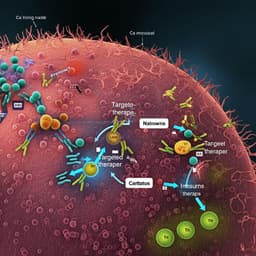
Advancements in clinical aspects of targeted therapy and immunotherapy in breast cancer
F. Ye, S. Dewanjee, et al.


