
Medicine and HealthMicrosystems & Nanoengineering
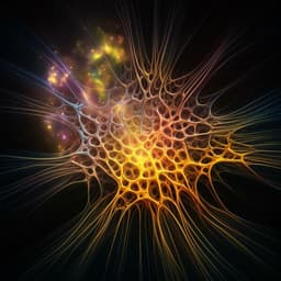
Infrared neural stimulation and inhibition using an implantable silicon photonic microdevice
Á. C. Horváth, S. Borbély, et al.
This groundbreaking study by Ágoston Csaba Horváth and colleagues presents a revolutionary multimodal photonic neural probe that not only controls temperature in deep brain tissue but also records the electrical responses of neurons. The innovative approach demonstrated the ability to enhance or suppress neuronal firing using infrared light—an advanced step in understanding thermally induced neural responses.
Related Publications
Explore these studies to deepen your understanding
Adjacent work that informs or extends this paper's methodology and findings.

Medicine and Health
Unprotected sidewalls of implantable silicon-based neural probes and conformal coating as a solution
P. Ghelich, N. F. Nolta, et al.

Education
Enhancing senior high school student engagement and academic performance using an inclusive and scalable inquiry-based program
L. D. Huyer, N. I. Callaghan, et al.
Medicine and Health
Common and distinct neural representations of aversive somatic and visceral stimulation in healthy individuals
L. V. Oudenhove, P. A. Kragel, et al.

Food Science and Technology
Synchronously Predicting Tea Polyphenol and Epigallocatechin Gallate in Tea Leaves Using Fourier Transform-Near-Infrared Spectroscopy and Machine Learning
S. Ye, H. Weng, et al.


