Medicine and Health
Infection with SARS-CoV-2 can cause pancreatic impairment
W. Deng, L. Bao, et al.
COVID-19 causes multi-organ complications, including pancreatic and endocrine involvement. Diabetes is a key risk factor for COVID-19 severity, with higher ICU admissions and mortality reported among diabetics. Experimental data indicate SARS-CoV-2 can infiltrate exocrine and endocrine pancreatic cells in vitro and in vivo, but whether pancreatic lesions are permanent, age-dependent, and whether direct β-cell infection occurs in patients of different ages remain unclear. Vaccines might also impact the pancreas; rare cases of post-vaccination acute pancreatitis have been reported, but whether vaccination induces lasting pancreatic or glycometabolic changes is unknown. Leveraging a repository of NHP specimens from 2020–2023, the study retrospectively examined pancreatic tissues from SARS-CoV-2-infected and vaccinated+infected rhesus macaques and compared them with human pancreatic autopsy samples. The goals were to map SARS-CoV-2 entry factor expression in pancreatic cell types, define viral infiltration across exocrine and endocrine compartments, quantify age-related pancreatic lesions (particularly islet damage) and glycometabolic alterations, and evaluate whether vaccination modifies glycemic homeostasis.
Prior clinical studies identified diabetes as a significant comorbidity associated with worse COVID-19 outcomes and increased ICU admissions. Reports suggested SARS-CoV-2 infects pancreatic exocrine and endocrine cells, though expression of entry factors (ACE2, TMPRSS2, NRP1) in β cells has been debated with contradictory findings. Case reports documented rare acute pancreatitis after COVID-19 vaccination. Mechanistic work implicated insulin resistance and infection of adipocytes and hepatocytes in COVID-19-related hyperglycemia. Islet amyloidosis is linked to type 2 diabetes pathophysiology and age-related changes. The study builds on this literature by systematically assessing in situ pancreatic infection, islet cell composition, fibrosis/amyloid, microvascular/stress markers, and multi-omics signatures across ages and vaccination states in NHPs and validating with human autopsy tissue.
Design: Retrospective analysis (2020–2023) of archived rhesus macaque (Macaca mulatta) pancreatic tissues and sera, supplemented with human pancreatic autopsy samples for comparison. Groups included: prototypic strain-infected adult NHPs (n=9), prototypic strain-infected elder NHPs (n=6), vaccinated+infected adult NHPs (n=35), and uninfected controls (adult n=3, elder n=3). Additional datasets encompassed 67 infected NHPs and 121 vaccinated+infected NHPs overall. Human samples: two COVID-19 autopsies and one non-diabetic elderly control pancreas. Viral strains and infection: Prototypic SARS-CoV-2/WH-09/human/2020/CHN; Delta-1/2021; Omicron-1/2021. Virus amplified on Vero cells; titers by TCID50. Infections assessed at 3 and 7 dpi (elder), and 7 dpi (adults); vaccinated cohorts challenged post-vaccination. Vaccination: Inactivated or subunit (recombinant protein) vaccines previously shown efficacious in NHPs; some analyses included longitudinal sampling 0–28 days post vaccination (dpv) without infection to isolate vaccine effects. Tissue processing and histopathology: Formalin-fixed paraffin-embedded pancreas sections (3–4 µm). H&E for general pathology; special stains: Congo red for amyloid, Masson’s trichrome for fibrosis. Quantification of amyloid/fibrosis areas as percent of tissue. Immunohistochemistry and multiplex immunofluorescence (mIF): Multi-label panels to detect SARS-CoV-2 S protein, islet hormones (insulin/β, glucagon/α, somatostatin/δ, PP), viral entry factors (ACE2, TMPRSS2, NRP1), markers of ductal (CK19), exocrine (trypsin), endocrine secretory granules (ICA512/PTPRN), insulin receptors (IRα, IRβ), microvascular/endothelial (CD31), fibrosis/stellate activation (COL1A1, α-SMA), proliferation (Ki67), inflammation/stress (ICAM-1, VCAM-1, G3BP1), and apoptosis (cleaved caspase-3). Antibodies validated; spectral imaging via Akoya Vectra Polaris; unmixing and quantification with inForm/Phenochart and ImageJ/GraphPad. Co-localization (e.g., S+ with endocrine markers) and double-positive cell percentages quantified per field (9–10 regions/slide; 3–4 slides/group). In situ hybridization (RNAscope): Detection of SARS-CoV-2 genomic RNA in FFPE sections (probe for S gene, positive-sense RNA). Serum biochemistry and immunoassays: Fasting samples measured for glucose, insulin, C-peptide, C-peptide/glucose ratio; GAD65 and GAD65 autoantibodies; PPP1R1A; amylase, lipase; ICAM-1, VCAM-1; glycated serum protein (GSP). Longitudinal 0–28 dpv profile for vaccinated-only NHPs. Electron microscopy: Ultrastructural assessment of islet cell organelles in elder infected animals (mitochondrial swelling, myeloid degeneration). Multi-omics: Serum quantitative proteomics, lipidomics, and metabolomics across cohorts: controls (n=3), elder infected 3 dpi (n=5), elder infected 7 dpi (n=5), adult infected prototypic 7 dpi (n=3), adult infected Delta 7 dpi (n=4), vaccinated+infected prototypic (n=3), vaccinated+infected Delta (n=4). Differential features identified, KEGG pathway enrichment (insulin resistance, lipid/atherosclerosis, pancreatic/insulin secretion, carbohydrate digestion/absorption, necroptosis, FcγR-mediated phagocytosis), PPI networks, and volcano plots generated. Ethics: Human sample use approved by relevant IRBs; NHP studies conducted in ABSL-3 with IACUC approval. Adult NHPs aged 3–5 years; elder 15–35 years.
- SARS-CoV-2 entry factors (ACE2, TMPRSS2, NRP1) were broadly expressed within β and α cells in NHP and human pancreas by mIF; in elder NHPs post-infection, NRP1 and TMPRSS2 increased, while ACE2 co-expression with insulin decreased.
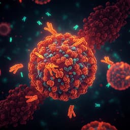
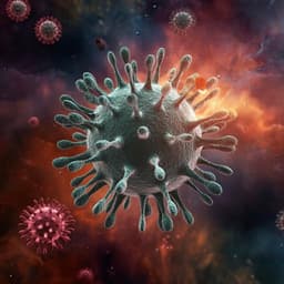
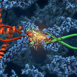
- Direct pancreatic infection: S protein and viral RNA detected in microvascular endothelium and dispersed across exocrine and endocrine cells. Co-staining confirmed S+ insulin β, S+ glucagon α, S+ somatostatin δ, and S+ PP cells in NHPs and humans.
- Elder NHP islet composition changes vs elder controls: α cells −63.49%, β cells −58.66%, δ cells −82.33%; PP cells increased. Human COVID-19 pancreas showed δ cells −72.47% vs controls; necrotic islets observed on H&E.
- Pathology severity: Adults showed mild pancreatic lesions; elders exhibited aggravated degeneration, amyloidosis, necrosis/atrophy of islets, ductal changes, and exocrine inflammation. Congo red–positive amyloid area increased by 5.86%; Masson’s-positive area by 4.16% in elder infected NHPs.
- Microvascular/stress markers in elder NHPs: ICAM-1 and G3BP1 significantly increased; VCAM-1 and cleaved caspase-3 not significantly changed. Increased ICAM1+β and G3BP1+β cell percentages; presence of glucagon+insulin+ double-positive cells suggests β-cell transdifferentiation.
- Fibrosis/stellate activation: In elder infected NHPs, COL1A1 (+23.98%), α-SMA (+14.97%), and CD31 (+21.14%) increased vs elder controls; insulin+COL1A1+ +2.24% and insulin+α-SMA+ +1.84%. Human COVID-19 pancreas: COL1A1 +19.52%, α-SMA +20.74%, Ki67 +5.78%; insulin+COL1A1+ +0.53%, insulin+α-SMA+ +0.69%; S+CD31+ cells 0.032.
- Glycometabolic profile (fasting): Adult infected NHPs had elevated C-peptide and C-peptide/glucose ratio (insulin resistance indicators); fasting glucose/insulin largely unchanged across groups. GAD65 antibodies were markedly elevated in adult infected NHPs; amylase/lipase not significantly changed; PPP1R1A notably elevated in one elder infected animal.
- Vaccination effects: In vaccinated+infected adults, insulin secretion not decreased; during 0–28 dpv without infection, GSP stable; insulin rose at 21 dpv (P=0.035) and 28 dpv (P=0.029); C-peptide, glucose, and ratio unchanged; GAD65 transient, nonsignificant rise; ICAM-1 slightly decreased. In situ, vaccination significantly increased IRα/IRβ expression and inhibited ICA512 vs control and infected groups, indicating activation of insulin signaling and preserved glucose homeostasis.
- Multi-omics: Adult infected NHPs showed enrichment of insulin resistance pathways and lipid/atherosclerosis signatures; pathways affecting pancreatic/insulin secretion and carbohydrate digestion/absorption also altered. Elder infected animals exhibited greater numbers of differentially expressed metabolites by 7 dpi (up to 440 vs control; 580 vs adult), with progressive metabolic dysfunction. Serum COL1A1 decreased from 3 to 7 dpi with correlated metabolite network changes; COL18A1 increased. Vaccinated+infected groups showed numerous differential proteins/lipids/metabolites but overall mild metabolic impact; notable increases in PE(O-20:0_20:5), CE(23:0), and N1-acetylspermidine as potential biomarkers.
The study demonstrates that SARS-CoV-2 directly infects pancreatic microvasculature and endocrine and exocrine cells in NHPs and humans. In adults, despite mild histopathology, infection is associated with insulin resistance signatures and elevated C-peptide and GAD65Ab, indicating β-cell stress or immune activation without overt hyperglycemia. This aligns with clinical observations that most acute COVID-19 patients do not develop frank β-cell failure, and suggests peripheral tissue insulin resistance (adipose, liver) may drive dysglycemia. In elder NHPs, infection markedly aggravates islet pathology, including amyloidosis, necrosis/atrophy, stellate activation and fibrosis, microvascular inflammation (ICAM-1), and cellular stress (G3BP1), with reductions in β-cell mass and insulin expression in situ. These pathologies mirror type 2 diabetes–like features and support an age-dependent vulnerability to post-COVID-19 glycometabolic dysfunction and potential diabetogenic effects. Proposed mechanisms include direct β-cell infection, endothelial injury with increased permeability and inflammatory infiltration, amyloid deposition constricting islet microenvironments, and stellate cell–driven matrix remodeling, collectively impairing β-cell function and survival. Vaccination, conversely, appears to preserve glycemic homeostasis by upregulating insulin receptor signaling (IRα/IRβ) and reducing ICA512 expression, with stable glycemic markers and only transient, nonsignificant autoantigen changes. The multi-omics findings integrate with histopathology, pinpointing insulin resistance, lipid dysregulation, and progressive metabolic derangement with age and infection duration.
SARS-CoV-2 infects pancreatic tissues and can impair endocrine function. Adult NHPs show insulin resistance and β-cell stress with mild pathology, while elder NHPs exhibit pronounced islet damage (amyloidosis, necrosis), stellate activation, fibrosis, β-cell loss, and worsened metabolic signatures, indicating an age-related risk for diabetes-like phenotypes post-infection. COVID-19 vaccination maintains insulin secretion homeostasis, likely via insulin receptor activation, and does not adversely alter glycemic markers. Clinically, heightened surveillance of glucose metabolism is warranted in elderly COVID-19 patients. Future studies should assess long-term and repeat infections, delineate mechanisms of β-cell injury and recovery, validate serum biomarkers (e.g., N1-acetylspermidine, lipid species), and expand human cohorts for translational confirmation.
- Retrospective design with limited and uneven serum sample sizes across groups, particularly in elders, which may underpower some comparisons (e.g., fasting insulin/glucose differences).
- Short-term infection window (primarily up to 7 days), limiting inference on long-term or recurrent infection effects on pancreatic structure/function.
- NHP models may not fully recapitulate human disease heterogeneity; apoptosis detection in islets was negative here, differing from some literature, potentially due to model/method differences.
- Human validation limited to a small number of autopsy cases and one control pancreas.
- Multi-omics based on serum may not capture tissue-compartmentalized changes; causality between specific molecular changes and pancreatic lesions cannot be definitively established.
Related Publications
Explore these studies to deepen your understanding of the subject.