Medicine and Health
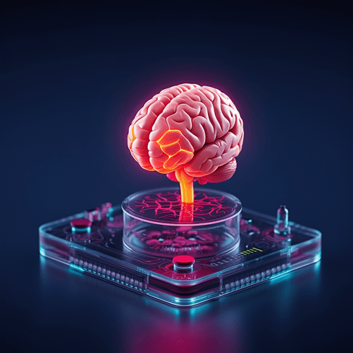
Human Brain Organoids-on-Chip: Advances, Challenges, and Perspectives for Preclinical Applications
H. Castiglione, P. Vigneron, et al.
Cerebral organoids coupled with microfluidic "brain-on-chip" systems promise reproducible, high-quality models of the human brain for disease modeling and drug discovery. This review — conducted by Héloïse Castiglione, Pierre-Antoine Vigneron, Camille Baquerre, Frank Yates, Jessica Rontard, and Thibault Honegger — explains how organoids-on-chips boost maturation, reproducibility, and compatibility with high-throughput pharmacological screens.
~3 min • Beginner • English
Introduction
Neurological disorders (e.g., Alzheimer’s disease, Parkinson’s disease, ALS, stroke, brain injuries) affect up to one billion people worldwide and are major causes of disability and mortality. Despite investment, CNS drug development has high failure rates, highlighting limitations of current experimental tools and the translational gap from animal models to humans. Brain complexity—cellular heterogeneity (neurons, glia, microglia, endothelial cells), vast synaptic networks, plasticity, immune privilege, and the blood–brain barrier—complicates predictive model development and drug delivery. Conventional 2D cultures lack 3D architecture and physiological microenvironments; 3D systems are more relevant but often irreproducible; microfluidic devices can recapitulate physiological environments under controlled flow. Pluripotent stem cell (PSC)-derived cerebral organoids can model aspects of human brain development and disease. Combining organoids with organ-on-chip (OoC) microfluidic systems (brain organoids-on-chips) offers improved physiological relevance and experimental control, potentially accelerating preclinical testing. The review clarifies nomenclature (neural vs. cerebral organoids) and outlines how microfluidic technology can address organoid limitations to support pharmacological applications.
Literature Review
The review synthesizes advances in cerebral organoids and their integration with microfluidics:
- Cerebral organoids (PSC-derived) recapitulate diverse brain regions (forebrain, midbrain, hindbrain, cortex, hippocampus, choroid plexus) and developmental processes (neurogenesis, migration, neuromorphogenesis, synaptogenesis), forming ventricular zone-like and subventricular zone-like layers with emergent cortical layering over months.
- Methodologies: Unguided protocols (e.g., Lancaster & Knoblich, 2013) rely on intrinsic patterning, generating heterogeneous organoids with multiple regional identities; regionalized (guided) protocols drive differentiation toward specific regions (cortex, midbrain, cerebellum, hippocampus, thalamus, hypothalamus), improving reproducibility at the cost of scope.
- Applications: Modeling neurodevelopmental disorders (microcephaly, Trisomy 21), prenatal toxicant exposures (nicotine, cadmium, cannabis, valproic acid), oncogenesis (engineered tumorigenesis), and aspects of neurodegenerative diseases (Alzheimer’s, Parkinson’s). Patient-derived iPSCs enable personalized disease modeling.
- Challenges limiting preclinical adoption: High inter- and intra-organoid heterogeneity, necrotic cores due to lack of vasculature (oxygen/nutrient diffusion limit ~400 µm), absence of microglia, incomplete late developmental features (gliogenesis, myelination), variability across protocols and undefined components (e.g., Matrigel).
- Microfluidic solutions: Controlled flow reduces shear stress, improves oxygenation and nutrient/waste exchange, can establish gradients, enhance differentiation and reproducibility, enable in situ analyses, and flexible designs (single/multi-chamber, co-cultures, compartmentalization, multi-organ chips).
Methodology
As a review, the paper systematically examines and categorizes brain organoid-on-chip architectures and protocols reported in the literature, detailing device designs, fabrication approaches, and organoid generation methods, along with their reported advantages and limitations for preclinical use:
- Device classifications: (i) microfluidic devices with 3D culture areas and channels (perfusable chambers and flow paths); (ii) devices with micropillar arrays enabling in situ EB formation and organoid growth; (iii) devices with integrated air–liquid interface (ALI) chambers to enhance oxygenation and size control.
- Protocols: Unguided (Lancaster’s protocol and variants) and region-specific guided differentiation (e.g., cortical organoids), with in situ vs. ex situ Matrigel embedding depending on device geometry.
- Comparative analysis: Summaries of cited studies include device dimensions (e.g., channel width/length, chamber diameters/heights), flow regimes (continuous perfusion vs. gravity-driven bidirectional flow), materials (optically clear microfluidics), and coupling with ECM (brain ECM-enriched Matrigel) or co-culture (endothelial/pericyte vascularization).
- Readouts surveyed: Structural (immunofluorescence, in-device imaging), functional (Ca2+ imaging, patch clamp, MEA), transcriptomics (RT-qPCR, RNAseq, single-cell/spatial approaches), viability/hypoxia (TUNEL, oxygen-sensing nanoparticles), and metabolite measurements (glucose, lactate; LC-MS).
- Industrial considerations: Standardization of organoid protocols (PSC source, feeder-free culture, passage/confluency controls, ECM alternatives), device manufacturing (commercial reproducible platforms, material absorption issues), HTS compatibility (non-invasive readouts, automation, data processing/algorithms), and complexification (BBB modeling, multi-organoid chips).
Key Findings
- Microfluidic perfusion devices (continuous flow via syringe pump) improved organoid viability and growth versus static culture; enabled modeling of prenatal nicotine exposure, revealing disrupted regionalization, neurite outgrowth, and premature differentiation (Wang et al. 2018; Lab Chip 2018; 33-day cultures).
- Devices modeling bidirectional cerebrospinal fluid-like flow (gravity-driven rocker, open chambers connected by microchannels) enhanced oxygen diffusion (PtTFPP-PUAN nanoparticles), reduced heterogeneity, increased neurogenesis/corticogenesis, and accelerated maturation (mature neurons/astrocytes/synapses; electrophysiological signals) by ~60 days; upper/deep cortical layer separation by ~120 days; effects amplified with human brain ECM-enriched Matrigel (Cho et al. 2021).
- Constrained-height chamber (150 µm) devices induced surface wrinkling/folding, enabling lissencephaly modeling (reduced convolutions in diseased organoids); supported in-device imaging and immunostaining, suggesting physical constraints can promote maturation (Karzbrun et al. 2018).
- Vascularized organoid-on-chip (central organoid chamber surrounded by endothelial/pericyte channels) established perfusable/permeable vascular networks and accelerated maturation at 15–30 days versus non-vascularized controls; potential BBB modeling scaffold (Salmon et al. 2022).
- Micropillar array platforms allowed fully in situ organoid generation (unguided or guided cortical), facilitating HTS-oriented workflows; used to model cadmium neurotoxicity (increased apoptosis, impaired differentiation/maturation) and prenatal VPA exposure (increased progenitors, inhibited neuronal differentiation, altered forebrain regionalization, autism-like features), and breast cancer-derived exosome impacts (impaired neurogenesis, activated oncogenic signaling) (Zhu/Yin; Cui et al.).
- Air–liquid interface (ALI) microfluidic chambers supported whole organoids with improved viability, reduced hypoxia, enhanced diameter homogeneity, and compatibility with standard multiwell formats (up to 169 organoids per 6-well); used to model prenatal cannabis exposure (reduced maturation, impaired neurite outgrowth, reduced firing rate) (Ao et al. 2020).
- Readouts and data handling: Most studies relied on IF and RT-qPCR/RNAseq; some incorporated Ca2+ imaging, patch-clamp, MEA (2D), oxygen and metabolite measurements; in-device imaging and RNA extraction feasible; non-invasive, automation-compatible readouts (medium sampling, MEA) highlighted for HTS.
- Industrial adoption pathways: Emphasis on standardization (PSC handling, ECM selection/alternatives), reproducible device manufacturing (considering material absorption), non-invasive monitoring (electrophysiology, metabolomics), and integration with BBB and multi-organoid chips.
Discussion
The synthesis indicates that integrating cerebral organoids with microfluidic platforms can mitigate key barriers to preclinical adoption: enhancing oxygenation and nutrient exchange, reducing heterogeneity, enabling controlled microenvironments and flow, and facilitating in situ analyses. These advances address the need for more predictive human-relevant models in CNS drug discovery, potentially improving translational outcomes compared to animal models or 2D cultures. However, routine pharmacological use requires:
- Standardized, reproducible organoid generation (preferably guided protocols for reduced heterogeneity), defined quality controls (PSC source/state, passage/confluency, EB morphology), and ECM strategies (moving beyond animal-derived Matrigel to defined hydrogels with appropriate mechanics).
- Scalable, reproducible device manufacturing with clear user protocols; materials chosen to minimize small-molecule absorption (e.g., PDMS considerations) and maintain optical/electrical compatibility.
- HTS-compatible, non-invasive readouts (electrophysiology via 3D MEA, medium-based biomarkers/metabolomics) with automated imaging/data pipelines and standardized biomarkers for viability, toxicity, and disease phenotypes.
- Model complexification for pharmacological relevance, including BBB incorporation for drug permeability and distribution, and multi-organoid/multi-organ systems for inter-tissue interactions.
Together, these strategies enhance experimental control and predictivity, aligning brain organoids-on-chips with pharmaceutical screening needs while reducing reliance on animal testing.
Conclusion
Cerebral organoids-on-chips synergistically combine the physiological complexity of 3D human brain models with the controllability of microfluidics, enabling more relevant preclinical testing environments (e.g., toxicology, delivery, screening) than conventional models. To achieve routine early-stage drug development use, the field must reduce variability and increase quality (maturation markers, shorter culture durations), establish standardized, reproducible protocols and device fabrication, and demonstrate translational predictive value alongside animal reduction. Once validated, two impactful directions are envisioned: personalized medicine using patient-derived cells and multi-organoid-on-chip platforms to capture inter-tissue interactions.
Limitations
- Intrinsic organoid heterogeneity (size, morphology, cell-type proportions, structural organization) within and between batches, especially with unguided protocols; intra-organoid heterogeneity and occasional non-neural germ layers or cystic cavities.
- Necrotic core formation due to lack of vasculature and diffusion limits (~400 µm), restricting size (~4–5 mm) and maturation; absence of microglia limits immune and maturation aspects.
- Incomplete recapitulation of late neurodevelopment (gliogenesis, myelination), cortical layering complexity, and gyrification in many models; long culture times (up to 120 days) for advanced features.
- Practical device constraints: Some formats require pumps or complex assembly, impede in situ Matrigel embedding, or lack individual organoid compartmentalization; limited control of flow in micropillar arrays; constrained devices may not scale industrially.
- Readout limitations: Predominant reliance on destructive IF/sectioning and RNA extraction; small medium volumes complicate reproducible metabolomics; surface-limited electrophysiology (2D MEA) misses deep layers.
- Materials issues: Absorption of small molecules (e.g., in PDMS) can confound drug testing; need for optically clear, electrically compatible materials with low adsorption.
- Standardization gaps: Variability in PSC culture (feeder-dependent vs. feeder-free), passage/confluency, EB morphology; undefined ECM components (Matrigel) with batch variability and supply constraints.
Related Publications
Explore these studies to deepen your understanding of the subject.

