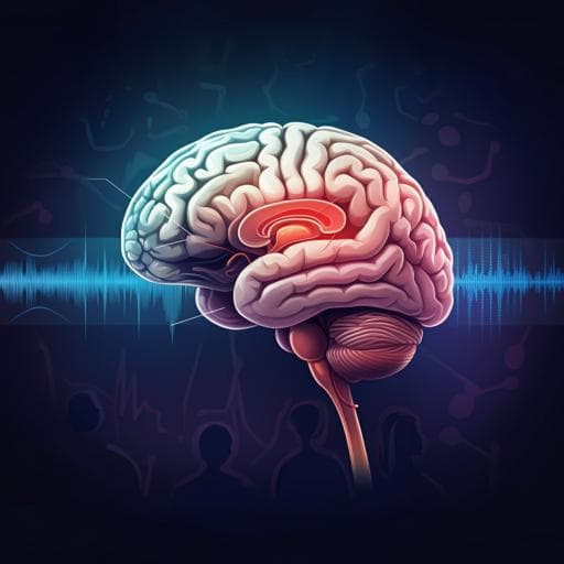
Highly demarcated structural alterations in the brain and impaired social incentive learning in *Tbx1* heterozygous mice
T. Hiramoto, A. Sumiyoshi, et al.
Explore these studies to deepen your understanding
Adjacent work that informs or extends this paper's methodology and findings.

Similar cognitive deficits in mice and humans in the chronic phase post-stroke identified using the touchscreen-based paired-associate learning task
W. Z. Chow, L. K. Ong, et al.

Evaluating trust and shared group identities in emergent social learning processes in the Zambezi river basin
C. K. Lumosi, C. Pahl-wostl, et al.

The impact of a social and emotional learning programme to improve pupils' educational inclusion in vocational education and training
F. D. Fernández-martín, I. Aznar-díaz, et al.
Gut microbiota determines the social behavior of mice and induces metabolic and inflammatory changes in their adipose tissue
O. Agranyomi, S. Meininger-mordechaï, et al.


