
Medicine and Health
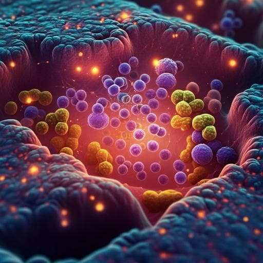
High resolution mapping of the tumor microenvironment using integrated single-cell, spatial and in situ analysis
A. Janesick, R. Shelansky, et al.
This groundbreaking study conducted by Amanda Janesick and colleagues from 10x Genomics Inc. reveals the potential of integrating cutting-edge single-cell and spatial technologies to provide new insights into breast cancer. By analyzing FFPE human breast cancer sections, they identify key molecular differences between tumor regions and highlight rare boundary cells that may play a role in cancer progression. Discover how this research could transform oncology diagnostics and therapeutics.
~3 min • Beginner • English
Introduction
The study addresses how to achieve high-resolution, spatially resolved characterization of the tumor microenvironment in FFPE breast cancer tissue by integrating complementary genomics technologies. Single-cell RNA profiling offers whole-transcriptome resolution but lacks spatial context, while spatial transcriptomics provides unbiased spatial information at spot-level resolution; high-plex in situ methods deliver subcellular spatial localization but are targeted and require careful integration. The authors hypothesize that combining whole-transcriptome single-cell (Chromium Single Cell Gene Expression Flex on FFPE), whole-transcriptome spatial (Visium CytAssist), and high-plex in situ (Xenium) will uncover molecular heterogeneity among DCIS and invasive regions, refine cell-type annotations, identify rare cell states (e.g., boundary cells), and reveal clinically relevant biomarkers (e.g., ER/PR/HER2) that are not apparent with any single modality. This integrative approach aims to improve understanding of DCIS-to-invasive transitions and support diagnostics and therapeutic development.
Literature Review
The paper situates the work within advances in single-cell genomics and spatial transcriptomics, noting large-scale efforts to catalog human cell types and methods to integrate single-cell and spatial data for transcript distribution prediction and deconvolution. It highlights the gap in definitively assigning transcripts to individual cells with spatial context at high gene plex and in analyzing high-resolution cell–cell and ligand–receptor interactions. Recently commercialized high-plex in situ platforms (CosMx, MERSCOPE, Molecular Cartography, Xenium) address subcellular resolution needs but pose integration challenges with whole-transcriptome datasets. Prior studies have linked specific markers (e.g., low KRT15) to prognosis and identified stromal and myoepithelial contributions to invasiveness; pathology concordance challenges underscore the need for molecularly informed spatial profiling. The study builds on these by integrating whole-transcriptome single-cell and spatial data with targeted in situ to refine cell states and spatial relationships in FFPE tissues.
Methodology
Design: Serial FFPE sections from human breast cancer blocks were profiled with three technologies. Sample #1 (Stage II-B, ER+/PR-/HER2+) provided scFFPE-seq, Visium CytAssist, and Xenium data; Sample #2 (ER−/PR−/HER2+) provided Xenium data for validation and discovery of boundary cells.
Single-cell (scFFPE-seq): FFPE curls (2×25 µm pooled to 50 µm) were dissociated (Miltenyi FFPE Tissue Dissociation Kit). ~600,000 cells prepared; 16,000 cells loaded per GEM well across four wells on Chromium X (target ~10,000 recovered cells per well). Libraries prepared with Chromium Single Cell Gene Expression Flex (RNA-templated ligation probe set; 18,536 genes targeted by 54,018 probes). Sequencing: Illumina NovaSeq, PE (28/10/10/90). Processing: Cell Ranger v7.0.1 (multi pipeline; GRCh38-2020-A). QC with scanpy (largest gene fraction ≤0.2; mitochondrial fraction ≤0.15; genes per cell ≥500). Dimensionality reduction with PCA (top 50 PCs) and t-SNE (monet v0.3.2). Differential gene expression (Loupe) using negative binomial exact or fast asymptotic beta tests.
Spatial (Visium CytAssist): 5 µm sections placed on glass slide, H&E stained, imaged, decoverslipped, decrosslinked, then transferred via CytAssist to Visium FFPE slides (0.42 cm² capture area). Libraries constructed per Visium FFPE protocol and sequenced (28/10/10/90). Processing: Space Ranger v2022.0705.1 (GRCh38-2020-A). Integration with scFFPE-seq facilitated by identical probe set. Spot deconvolution: Robust Cell Type Decomposition (RCTD; spacexr 2.0) using scFFPE-seq reference. Visium–Xenium registration via H&E alignment with manually defined keypoints and RANSAC homography.
In situ (Xenium): 5 µm FFPE sections on Xenium slide; deparaffinization and permeabilization. Targeted panel of 313 genes (Xenium Human Breast Panel, 280 core genes + 33 add-ons), curated from human breast single-cell atlases. Probe design with two target-hybridizing arms and gene-specific barcode; ligation to circular DNA, followed by rolling circle amplification (RCA). Hybridization: 50 °C overnight (10 nM); ligation at 37 °C; amplification (1 h at 4 °C, then 2 h at 37 °C). Imaging on Xenium Analyzer (12×24 mm area per slide; wide-field epifluorescence; 15 cycles of fluorescent probe hybridization/imaging/removal; Z-stacks with 0.75 µm step). Image preprocessing: lens distortion correction, stitching (DAPI), subsampling Z to 3 µm for segmentation. RCA puncta detection via Gaussian fitting; global maximum-likelihood decoding against a codebook across cycles/channels. Quality filtering: Phred-style Q-Score calibration using negative control codewords/probes and unassigned codewords; transcripts with Q ≥ 20 retained. Cell segmentation: DAPI-based deep neural network approach similar to CellPose; 2D segmentation from combined Z-planes; nuclei expansion assigns transcripts to nearest nucleus within 15 µm, preventing overlaps; trained to F1 > 0.80 at IoU 0.5. Outputs: feature–cell matrices (HDF5/MEX), per-transcript coordinates, cell boundaries CSV.
Supervised and unsupervised labeling: scFFPE-seq clusters annotated by DGE and literature; labels transferred to Xenium via nearest neighbors in PC space (30-NN; ≥50% agreement to assign; otherwise unlabeled). Unsupervised clustering on Xenium Sample #2 with Seurat 5 (development branch), resolution 0.3; subclustering for epithelial and macrophage subsets; visualization with ImageFeaturePlot and custom ggplot2 transcript overlays.
Integration and analytics: Registration of Xenium to Visium to compute Xenium pseudobulk counts within Visium spots (spot interpolation by proximity). DGE for ROIs with scanpy (Wilcoxon on log-normalized counts). Whole-transcriptome DGE in Visium for triple-positive region vs other malignant spots (Loupe; multiple-testing correction via Benjamini–Hochberg). Gene ontology enrichment (Enrichr; BioPlanet, Reactome). Benchmarking sensitivity across platforms: median gene sensitivity comparisons at matched depths (~10,000 reads/cell for scFFPE-seq; 20,000 for 3'/5' GEX) and panel-matched downsampling; gene dropout Venn analysis. IF validation post-Xenium with HER2 and CD20 antibodies; registration via SIFT (cv2).
Key Findings
- Discovery and atlas: scFFPE-seq from Sample #1 yielded 17 clusters (t-SNE) with a median of 1480 genes per cell. Visium identified 17 spatial clusters with a median of 5712 genes per spot, localizing two molecularly distinct DCIS regions (DCIS #1, DCIS #2) and invasive tumor, plus immune, stromal, adipocyte territories.
- Xenium profiling: In one section, 167,885 cells and 36,944,521 total transcripts (Q ≥ 20) were detected, with median 166 transcripts per cell (10th percentile 61; 90th percentile 372). With a 313-gene panel, Xenium detected a median of 62 genes per cell vs scFFPE-seq downsampled to the same genes detecting a median of 34 genes per cell. Negative control probes constituted 0.026% and decoding controls 0.01% of counts, indicating high specificity. Replicate serial sections showed highly correlated transcript counts (r² = 0.99) and similar cell-type proportions.
- Labeling and segmentation: Supervised label transfer from scFFPE-seq to Xenium unambiguously labeled 86% of Xenium cells. Unsupervised labeling on Xenium recovered major cell types but could not fully resolve DCIS subtypes or proliferative tumor cells, underscoring the benefit of integration and accurate segmentation.
- Sensitivity and concordance benchmarking: At matched sequencing depths (~10k reads/cell), scFFPE-seq median gene sensitivity exceeded Chromium 3' and 5' GEX (fresh/frozen). Xenium sensitivity (transcripts per cell) was comparable to scFFPE-seq (UMIs per cell) when downsampled to the 313-gene panel. In the shared capture area (78% of Visium section), Visium showed 3.6× more total transcripts (whole-transcriptome vs targeted), yet Xenium exhibited 8.4× higher median gene sensitivity across panel genes. Spatial expression concordance was strong (e.g., TACSTD2, r² = 0.88 between platforms). IF protein/RNA overlays (HER2, CD20) matched spatially on the same section.
- Regional heterogeneity: Three ROIs (DCIS #1, DCIS #2, invasive) showed distinct compositions: ACTA2+ myoepithelial cells were prominent in DCIS #2, less in DCIS #1, and absent in invasive ROI; invasive tumor cells were present within DCIS #2; endothelial cells were slightly enriched in the invasive ROI. DCIS #2 exhibited fewer KRT15+ myoepithelial cells and more invasive markers than DCIS #1, consistent with higher invasiveness.
- Molecular markers: DCIS #1 featured ALDH1A3, KRT15, KRT23 in myoepithelial cells; DCIS #2 had GJB2+ stromal cells; MZB1 marked DCIS #1; macrophage marker MMP12 was absent from the invasive ROI. AGR3 marked tumor epithelium in DCIS but not invasive regions.
- Triple-receptor-positive niche: Despite overall HER2+/ER+/PR− annotation, Xenium revealed a small DCIS region triple positive for ERBB2/ESR1/PGR, adjacent to adipocytes and lacking a KRT15+ myoepithelial layer. scFFPE-seq detected few PGR+ cells without coexpression of ERBB2 or ESR1. Integration via spot interpolation identified 5–6 Visium spots covering this niche; whole-transcriptome DGE uncovered 48 DEGs vs PGR− DCIS #1 and 44 DEGs vs PGR− DCIS #2 (log2FC > 1.5; p < 0.05). GO terms in the triple-positive region included ErbB4 and estrogen receptor signaling; DCIS #1 enriched for metabolic pathways; DCIS #2 for interferon signaling.
- Boundary cells: In Sample #2, Xenium identified a rare population at deteriorating myoepithelial boundaries coexpressing tumor (ERBB2, ABCC11) and myoepithelial markers (MYLK, DST). Controls in normal ducts showed minimal transcript commingling, supporting specificity. Projecting this boundary-cell signature onto scFFPE-seq (Sample #1) identified ~1% of cells with the profile (~283 cells). DGE highlighted boundary-cell-enriched genes CX3CL1, CCL28, PROM1, and KLK5 not elevated in tumor or myoepithelial cells.
- Practical pathology insights: Xenium detected ducts with normal morphology containing tumor markers, suggesting potential for earlier molecular detection prior to overt histological changes.
Discussion
By integrating whole-transcriptome single-cell and spatial data with high-plex in situ profiling on FFPE sections, the study overcomes limitations of each modality alone. scFFPE-seq sensitively defines cell types and states but lacks spatial context; Visium maps tissue architecture and aids in identifying spatially distinct tumor subtypes but has spot-level resolution; Xenium assigns transcripts to individual cells with subcellular localization yet is targeted. Together, they reveal fine-grained heterogeneity within DCIS and invasive regions, delineate myoepithelial diversity and loss at invasive fronts, identify small triple-receptor-positive niches missed by discovery-only modalities, and uncover rare boundary cells potentially involved in DCIS-to-invasive transitions. The strong cross-platform concordance and IF validation underscore assay specificity and robustness. These findings address the core question of resolving tumor microenvironment complexity in FFPE tissues and demonstrate clinical relevance by refining biomarker landscapes (ER/PR/HER2, AGR3) and highlighting molecular changes preceding morphological invasion, which could inform prognosis and treatment stratification.
Conclusion
This work demonstrates that integrating Chromium scFFPE-seq, Visium CytAssist, and Xenium in situ on FFPE breast cancer sections provides high-resolution, spatially resolved characterization of the tumor microenvironment. The approach maps distinct DCIS subtypes and invasive regions, refines myoepithelial and immune landscapes, identifies a rare triple-receptor-positive DCIS niche, and discovers boundary cells coexpressing tumor and myoepithelial markers with unique gene signatures (CX3CL1, CCL28, PROM1, KLK5). Cross-platform benchmarking shows high sensitivity, specificity, and reproducibility, with complementary strengths across modalities. Future directions include validating boundary-cell roles in invasion across larger cohorts, expanding targeted panels or leveraging whole-transcriptome in situ, integrating additional modalities (e.g., imaging mass cytometry, CITE-seq), and translating the workflow to broader FFPE biobanks for translational and clinical research.
Limitations
- Sampling and replication: For Sample #1, scFFPE-seq was N=1 due to material constraints from using a single FFPE block across multiple technologies, potentially limiting generalizability. Xenium and Visium were run on two serial sections each, but biological replicates across donors were limited.
- Targeted panel constraints: Xenium used a 313-gene targeted panel, which, while capturing major heterogeneity, cannot discover markers outside the panel and was less resolutive for some subtypes (e.g., DCIS subclasses via unsupervised clustering).
- Segmentation and proximity effects: Accurate cell assignment near tightly apposed cell types (e.g., immune–stromal interfaces, adipocytes) is challenging; adipocytes were difficult to segment due to lipid-rich morphology and did not form distinct clusters.
- Spatial resolution of Visium: Spot-level resolution mixes cell types, necessitating deconvolution and integration; some Visium clusters remained mixed/unannotated.
- Pathology and registration dependencies: ROI definitions and interpretations rely on histology, registration accuracy, and expert annotations; small registration errors could affect spot interpolation and deconvolution.
- Cohort and tissue type: Findings are from FFPE breast cancer specimens; broader validation across cancers and sample processing types (fresh/frozen) is needed.
Related Publications
Explore these studies to deepen your understanding of the subject.