Medicine and HealthNature Communications
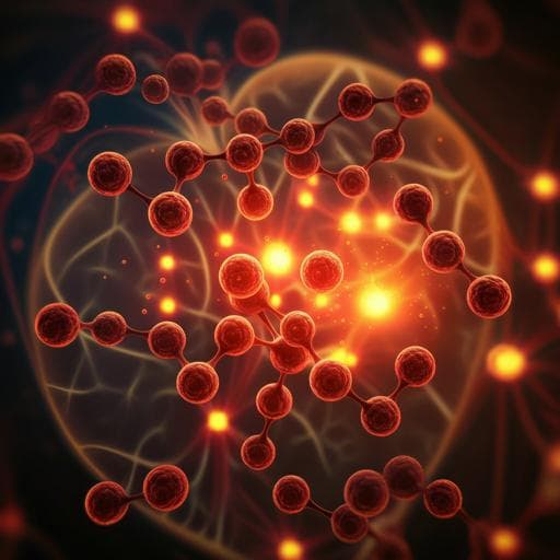
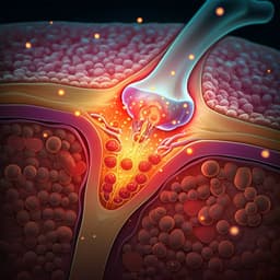
Electrophysiological engineering of heart-derived cells with calcium-dependent potassium channels improves cell therapy efficacy for cardioprotection
P. Vigneault, S. Parent, et al.
This innovative research by Patrick Vigneault and colleagues explores how calcium-activated potassium channels could revolutionize cell therapy for heart diseases. The study highlights the potent role of KCa3.1 in enhancing heart function and cell growth, offering promising avenues for cardiac protection and regeneration.
Related Publications
Explore these studies to deepen your understanding
Adjacent work that informs or extends this paper's methodology and findings.

Medicine and Health
Neutralizing IFNγ Improves Safety without Compromising Efficacy of CAR-T Cell Therapy in B-cell Malignancies
S. Manni, F. D. Bufalo, et al.

Food Science and Technology
Cell-based, cell-cultured, cell-cultivated, cultured, or cultivated. What is the best name for meat, poultry, and seafood made directly from the cells of animals?
W. K. Hallman, W. K. H. Ii, et al.
Medicine and Health
Mesenchymal stem cell-derived exosomes as a promising cell-free therapy for knee osteoarthritis
D. Luo, H. Zhu, et al.
Medicine and Health
Orally desensitized mast cells form a regulatory network with Treg cells for the control of food allergy
Y. Takasato, Y. Kurashima, et al.


