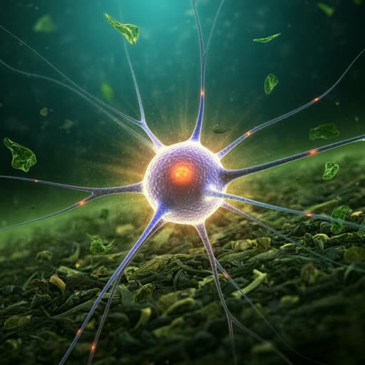
EGCG Attenuates CA1 Neuronal Death by Regulating GPx1, NF-κB S536 Phosphorylation and Mitochondrial Dynamics in the Rat Hippocampus following Status Epilepticus
A. V. Kozlov, S. Javadov, et al.
Explore these studies to deepen your understanding
Adjacent work that informs or extends this paper's methodology and findings.

Mitochondrial Oxidative Stress Is the General Reason for Apoptosis Induced by Different-Valence Heavy Metals in Cells and Mitochondria
S. M. Korotkov
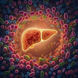
Stigmasterol attenuates hepatic steatosis in rats by strengthening the intestinal barrier and improving bile acid metabolism
Y. Zhang, Y. Gu, et al.

Dopamine release and dopamine-related gene expression in the amygdala are modulated by the gastrin-releasing peptide in opposite directions during stress-enhanced fear learning and extinction
Y. Morishita, I. Fuentes, et al.

Dopamine release and dopamine-related gene expression in the amygdala are modulated by the gastrin-releasing peptide in opposite directions during stress-enhanced fear learning and extinction
Y. Morishita, I. Fuentes, et al.


