
Chemistry
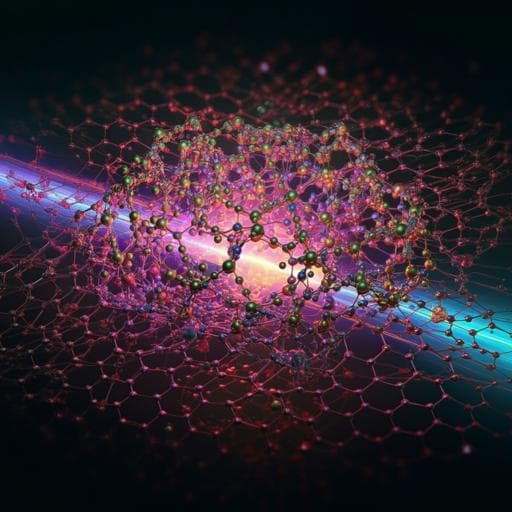
Dynamic three-dimensional structures of a metal-organic framework captured with femtosecond serial crystallography
J. Kang, Y. Lee, et al.
This groundbreaking study leverages time-resolved serial femtosecond crystallography (TR-SFX) to explore the intricate structural dynamics of a metal-organic framework involving Fe porphyrins and hexazirconium nodes. The research, conducted by a team of experts, uncovers unique structural pathways that reveal the exciting potential of TR-SFX for unraveling chemical systems at the atomic level.
~3 min • Beginner • English
Introduction
The study addresses whether femtosecond time-resolved serial femtosecond crystallography (TR-SFX), previously successful for proteins, can be extended to resolve ultrafast structural dynamics in non-biological crystalline materials composed of small-molecule building blocks. Static crystallography and low-temperature photocrystallography capture snapshots before/after stimuli but lack direct real-time 3D electron density evolution, and femtosecond X-ray/electron diffraction with few Bragg spots cannot reconstruct full electron density maps. The authors target a porphyrinic Zr-based MOF, PCN-224(Fe)-CO, which undergoes photoinduced CO dissociation, to visualize in real time how structural perturbations at Fe porphyrin propagate to Zr6 nodes. The work is significant because small-molecule crystals present challenges (smaller lattice constants, sparse Bragg peaks, stronger absorption and weak photoresponses) that have prevented TR-SFX from being applied to such materials.
Literature Review
Prior crystallographic approaches at synchrotrons offer ~100 ps resolution and are limited for irreversible, slow-recovery, or sub-100 ps processes. Femtosecond X-ray and electron diffraction capturing a few Bragg reflections provide partial insights but not full 3D electron density maps. TR-SFX has revealed ultrafast structural dynamics in proteins leveraging XFEL sources, whereas small-molecule crystalline systems and porous frameworks have not been investigated with TR-SFX; only static SFX on chemical crystals has been reported. Light-initiated dynamics in MOFs have been studied by diverse time-resolved spectroscopies, highlighting rich photophysics but lacking time-resolved crystallographic visualization. Technical obstacles in small-molecule crystals include sparse Bragg peaks due to small unit cells and limited optical penetration causing weak signals.
Methodology
- System: Porphyrinic Zr MOF PCN-224(Fe) with FeTCPP linkers and Zr6 nodes, CO-ligated to form PCN-224(Fe)-CO to enable photoinduced CO dissociation.
- Sample preparation: Micro-sized single crystals (10–20 µm) synthesized and activated. Crystals were mounted on a custom fixed-target holder sealed under CO to maintain ligation. Fixed-target delivery minimizes sample consumption and avoids solvent/medium compatibility issues.
- TR-SFX setup: Pump–probe at PAL-XFEL XSS beamline. Pump: 400 nm, <100 fs pulses; Probe: 14.5 keV XFEL pulses, ~11 µm FWHM; laser beam ~45 µm FWHM, fluence ~5 mJ mm^-2; beams crossed at 10°. High X-ray energy increased the number of measurable Bragg peaks for the small unit cell.
- Data collection: 33 pump–probe delays from –3.9 ps to 3 ns; >15,000 images per delay; Rayonix MX225-HS detector (4×4 binning). Fixed-target raster scanning ensured fresh crystals for each shot.
- Data processing: Hit finding with Cheetah; indexing/integration with CrystFEL (XGANDALF), using ‘rings-sat-cen’ integration; merging with partialator. High-resolution data beyond 0.9 Å excluded due to noise. Space group determination with XPREP. STRUCTURE solution with SHELXT and refinement with SHELXL (full-matrix least squares on F^2; non-H atoms anisotropic; H isotropic). Residual electron density in pores handled with PLATON SQUEEZE; Zr-cluster disorder modeled.
- Electron density maps: Reference electron density and structure from negative delays (–3.9, –2.4, –0.6, –0.2 ps). Difference structure factor amplitudes ΔF obtained by subtracting pre-excited from each time delay; DED maps generated using ground-state phases.
- SVD and kinetics: Regional DED maps around Zr node and FeCO site decomposed by SVD to obtain time-invariant spatial components (LSVs) and time profiles (RSVs). The dominant component corresponds to vibrational heating (validated by Wilson B-factor analysis). Secondary components capture anisotropic, coherent motions. Global fits of key RSVs used a Gaussian IRF (200 fs FWHM) and a combination of exponential decay and damped cosine, yielding shared time constants.
- Kinetic model and SADED: A three-species kinetic model extracted species-associated DED (SADED) maps: Iosc (coherent oscillation), It (transient), Ihot (vibrationally hot). Full-unit-cell DED maps were used for decomposition.
- Extrapolated maps and structural refinement: Species-associated difference structure factors from SADEDs combined with ground-state structure factors using photoconversion yields p (Ihot=1.0, It=0.20, Iosc=0.25) to form extrapolated structure factors corresponding to pure species. Intensities (|Fextr|^2) were refined to obtain atomic models and key structural parameters (Fe doming; Zr–Zr distances).
Key Findings
- TR-SFX captured three distinct structural pathways in PCN-224(Fe)-CO following 400 nm excitation and CO dissociation:
1) Coherent oscillatory species (Iosc): Period 5.55 ± 0.01 ps (frequency ~6.01 cm^-1 or 0.18 THz) with damping time constant 2.68 ± 0.02 ps. Anisotropic DED features: along the Zr positional disorder d-axis and Fe doming/undoming perpendicular to the porphyrin plane. CO shows negative density consistent with dissociation. From extrapolated maps: Zr–Zr distance 1.057(7) Å (+0.216 Å vs ground). Fe doming exhibits extremes approximately +0.443 Å and –0.312 Å relative to the ground-state position.
2) Transient species (It): Forms within the instrument response (<200 fs) and decays with 47.1 ± 0.5 ps. Shows enhanced Zr disorder along d-axis and a characteristic Fe motion along the Fe–CO axis (negative density at original Fe position; positive lobes above/below). From refinement: Fe doming 0.716(7) Å (+0.119 Å); Zr–Zr distance 0.907(10) Å (+0.066 Å).
3) Vibrationally hot species (Ihot): Isotropic broadening around Fe, Zr, and CO positions; dominates at long delays up to 3 ns. Thermal rise bi-exponential with 1.143 ± 0.005 ps and 11.32 ± 0.07 ps. From refinement: Fe doming 0.684(4) Å (+0.087 Å); Zr–Zr distance 0.891(7) Å (+0.050 Å).
- Ground-state parameters (for comparison): Fe doming 0.597(6) Å; Zr–Zr distance 0.841(11) Å.
- Photoconversion yields used for extrapolated-map analysis: Iosc ~25%, It ~20%, Ihot 100%.
- DED features evolve from anisotropic (early time) to isotropic (late time), consistent with transition from coherent, directed motions to vibrational heating.
- The coherent low-frequency oscillation suggests a phonon mode in the solid MOF, with mechanical coupling between the photoactive Fe porphyrin and remote Zr nodes via the linker framework.
- High-quality ED maps obtained across 33 delays validated ground-state structure and enabled SVD-based kinetic separation and structural refinement; R1 factors for species were comparable to or lower than static structures (e.g., Ihot 7.74%).
Discussion
The study demonstrates that TR-SFX can recover time-resolved 3D electron density changes in a non-protein crystalline material, addressing the long-standing challenge of capturing ultrafast structural dynamics in small-molecule crystals. The results reveal rapid, organized anisotropic motions initiated by CO photodissociation at Fe porphyrin sites that propagate coherently to Zr nodes, evidencing mechanical coupling across the MOF framework. The observed 0.18 THz oscillation aligns with a low-frequency phonon mode, while the subsequent rise of a vibrationally hot, isotropic component quantifies energy flow and lattice heating.
By separating species-associated DEDs and refining extrapolated maps, the work links kinetics to concrete structural parameters: changes in Fe doming correlate positively with Zr–Zr distances across species, indicating concerted framework response. Compared with protein TR-SFX, the pronounced vibrationally hot contribution here underscores differences in heat dissipation (MOFs lacking extensive solvent/buffer), providing insights into energy management in porous solids. Overall, the findings confirm that TR-SFX can dissect coherent, transient, and thermal components of structural dynamics in MOFs, advancing understanding of photoinduced processes in functional crystalline materials.
Conclusion
This work extends TR-SFX beyond biomacromolecules to a metal-organic framework, PCN-224(Fe)-CO, resolving ultrafast, three-dimensional structural dynamics with femtosecond timing. Three structural pathways were extracted and structurally refined: a coherent oscillation (5.55 ps period, 2.68 ps damping), a transient intermediate (47.1 ps decay), and a vibrationally hot species with ps-scale rise. The approach quantified Fe doming and Zr–Zr distance changes, revealing their positive correlation and the phonon-like coupling between porphyrin and Zr nodes.
The study establishes TR-SFX as a viable probe of ultrafast structural changes in porous materials and small-molecule crystals. Future directions include applying TR-SFX to diverse MOFs and covalent frameworks, exploring other stimuli (e.g., redox, pressure, guest adsorption), mapping energy flow across frameworks, and integrating computation to assign phonon modes and refine kinetic models.
Limitations
- Sparse Bragg peaks from the small unit cell required high X-ray energy (14.5 keV) to obtain sufficient reflections; data beyond 0.9 Å were excluded due to noise.
- Small-molecule crystals have higher optical absorption, limiting pump penetration and reducing photoinduced signal strength, complicating analysis.
- The Zr cluster exhibits intrinsic positional disorder along the d-axis, necessitating disorder modeling; residual electron density in pores required PLATON SQUEEZE and may include contributions from disordered solvent/gas or partial MOF-525 occupancy.
- Instrument response function was approximated as a 200 fs FWHM Gaussian; kinetic extraction assumes convolution with this IRF and a specific kinetic form (exponential and damped cosine).
- Photoconversion yields for species in extrapolated-map refinement were estimated (Iosc ~25%, It ~20%, Ihot 100%), and linear extrapolation was used to obtain pure-species structure factors, which introduces model dependence.
- No absorption correction was applied (estimated negligible for 10–20 µm crystals), but residual systematic errors cannot be fully excluded.
Related Publications
Explore these studies to deepen your understanding of the subject.