Food Science and TechnologyThe ISME Journal
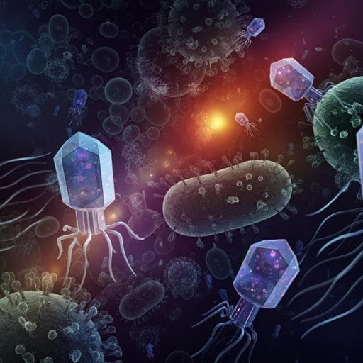
Dominance of phage particles carrying antibiotic resistance genes in the viromes of retail food sources
P. Blanco-picazo, S. Morales-cortes, et al.
Explore the exciting findings of a study that uncovers the significant roles of phages, outer membrane vesicles, and contaminating DNA in antibiotic resistance genes within food viromes. Conducted by a team of researchers including Pedro Blanco-Picazo and Sara Morales-Cortes, this research reveals the intricate dynamics of viromes in our food systems.
Related Publications
Explore these studies to deepen your understanding
Adjacent work that informs or extends this paper's methodology and findings.
Environmental Studies and Forestry
Seasonality impels the antibiotic resistance in Kelani River of the emerging economy of Sri Lanka
M. Kumar, G. G. T. Chaminda, et al.
Veterinary Science
Virulence-determinants and antibiotic-resistance genes of MDR- *E. coli* isolated from secondary infections following FMD-outbreak in cattle
A. M. Algammal, H. F. Hetta, et al.

Medicine and Health
Antidepressants promote the spread of extracellular antibiotic resistance genes via transformation
J. Lu, P. Ding, et al.

Social Work
Seeds and the city: a review of municipal home food gardening programs in Canada in response to the COVID-19 pandemic
J. Music, L. Mullins, et al.


