Medicine and HealthLight: Science & Applications
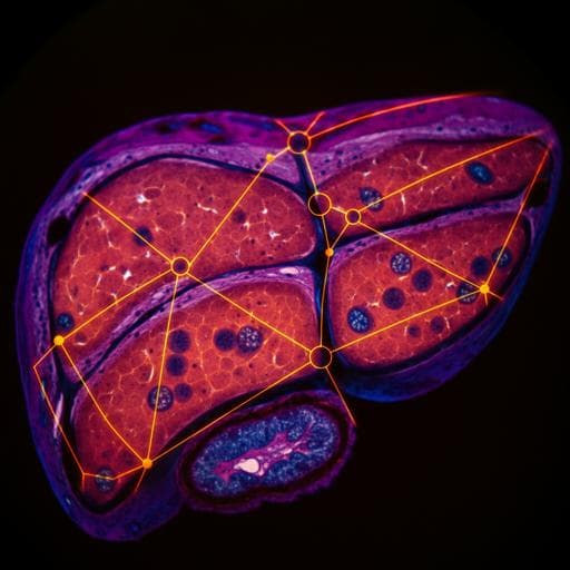
Deep learning-based virtual staining, segmentation, and classification in label-free photoacoustic histology of human specimens
C. Yoon, E. Park, et al.
Discover a groundbreaking deep learning framework for automated virtual staining, segmentation, and classification in label-free photoacoustic histology of human specimens. Researchers Chiho Yoon, Eunwoo Park, Sampa Misra, Jin Young Kim, Jin Woo Baik, Kwang Gi Kim, Chan Kwon Jung, and Chulhong Kim achieved remarkable accuracy in classifying liver cancers, establishing new potential for digital pathology.
Related Publications
Explore these studies to deepen your understanding
Adjacent work that informs or extends this paper's methodology and findings.
Medicine and Health
Deep learning-based virtual H&E staining from label-free autofluorescence lifetime images
Q. Wang, A. R. Akram, et al.
Medicine and Health
Automated detection of intracranial aneurysms using skeleton-based 3D patches, semantic segmentation, and auxiliary classification for overcoming data imbalance in brain TOF-MRA
S. Ham, J. Seo, et al.

Computer Science
A comprehensive review of deep learning in EEG-based emotion recognition: classifications, trends, and practical implications
W. Ma, Y. Zheng, et al.

Medicine and Health
Recent Advancements and Perspectives in the Diagnosis of Skin Diseases Using Machine Learning and Deep Learning: A Review
J. Zhang, F. Zhong, et al.


