Medicine and Healthnpj Imaging
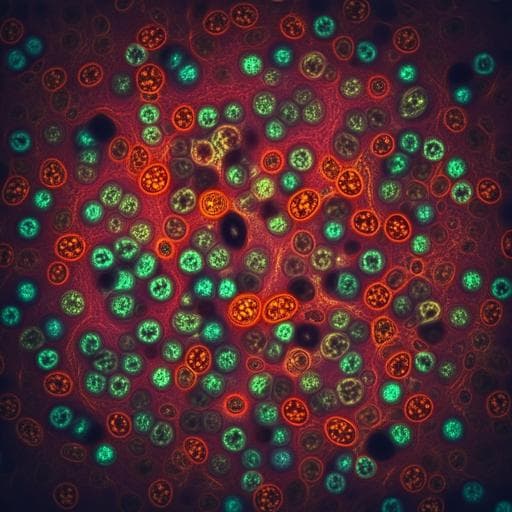
Deep learning-based virtual H&E staining from label-free autofluorescence lifetime images
Q. Wang, A. R. Akram, et al.
This groundbreaking research by Qiang Wang, Ahsan R. Akram, David A. Dorward, Sophie Talas, Basil Monks, Chee Thum, James R. Hopgood, Malihe Javid, and Marta Vallejos introduces a deep learning approach for producing virtual H&E stained images from label-free FLIM images. The results showcase improved accuracy in interpreting cellular structures, potentially revolutionizing tissue histology in cancer diagnostics.
Related Publications
Explore these studies to deepen your understanding
Adjacent work that informs or extends this paper's methodology and findings.
Medicine and Health
Deep learning-based virtual staining, segmentation, and classification in label-free photoacoustic histology of human specimens
C. Yoon, E. Park, et al.
Biology
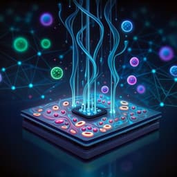
COSMOS: a platform for real-time morphology-based, label-free cell sorting using deep learning
M. Salek, N. Li, et al.
Medicine and Health
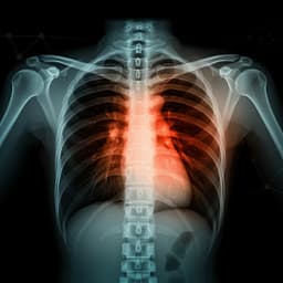
COVID-19 Prognosis from Chest X-ray Images by using Deep Learning Approaches: A Next Generation Diagnostic Tool
M. Pal, S. Parij, et al.
Medicine and Health
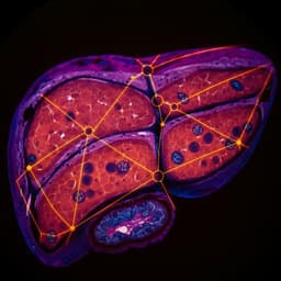
Using deep learning to predict abdominal age from liver and pancreas magnetic resonance images
A. L. Goallec, S. Diai, et al.


