
BiologyNature Structural & Molecular Biology
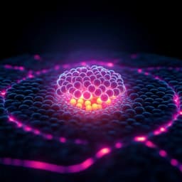
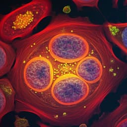
Cryo-electron tomography reveals how COPII assembles on cargo-containing membranes
E. Pyle, E. A. Miller, et al.
This research conducted by Euan Pyle, Elizabeth A. Miller, and Giulia Zanetti delves into the intriguing world of COPII coat architecture, revealing how these proteins assemble into spherical vesicles on membranes. Using cutting-edge cryo-electron tomography, the study uncovers the fascinating details behind membrane curvature generation and the structural dynamics of COPII transport carriers.
Related Publications
Explore these studies to deepen your understanding
Adjacent work that informs or extends this paper's methodology and findings.

Biology
Streamlined structure determination by cryo-electron tomography and subtomogram averaging using TomoBEAR
N. Balyschew, A. Yushkevich, et al.

Social Work
How the crisis of trust in experts occurs on social media in China? Multiple-case analysis based on data mining
Y. Wen, X. Zhao, et al.
Engineering and Technology
On-chip ultrasensitive and rapid hydrogen sensing based on plasmon-induced hot electron-molecule interaction
L. Wen, Z. Sun, et al.
Biology
High-dimensional super-resolution imaging reveals heterogeneity and dynamics of subcellular lipid membranes
K. Zhanghao, W. Liu, et al.


