Health and FitnessScientific Reports
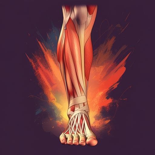
Considerations on the human Achilles tendon moment arm for in vivo triceps surae muscle-tendon unit force estimates
D. Holzer, F. K. Paternoster, et al.
This study, conducted by Denis Holzer and colleagues, explores how varying Achilles tendon moment arm-angle functions impact the force-angle relationship of the triceps surae muscle-tendon unit during maximum plantarflexion. The findings reveal that these functions significantly affect force estimations, emphasizing the need for individualized approaches in biomechanical research.
Related Publications
Explore these studies to deepen your understanding
Adjacent work that informs or extends this paper's methodology and findings.

Biology
Revised Estimates for the Number of Human and Bacteria Cells in the Body
R. Sender, S. Fuchs, et al.

Computer Science
On Multimodal Emotion Recognition for Human-Chatbot Interaction in the Wild
N. Kovačević, M. Gross, et al.

Education
Not-for-profit or for-profit? Research on the high-quality development path of private universities in China based on system dynamics
S. Duan, H. Yang, et al.

Social Work
Democratizing the discourse on criminal justice in social media: the activity for justice for Roman Zadorov as a case study
A. Lev-on


