Medicine and Health
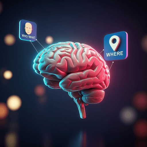
Concept and location neurons in the human brain provide the 'what' and 'where' in memory formation
S. Mackay, T. P. Reber, et al.
Our brains bind the 'who/what' and 'where' of experiences into episodic memories. By recording single-neuron activity in the human medial temporal lobe during item-location encoding, this study identifies two specialized neuron groups—concept cells in hippocampus/amygdala/entorhinal cortex and parahippocampal location-selective neurons—whose heightened firing predicts successful memory formation. Research conducted by Authors present in <Authors> tag.
~3 min • Beginner • English
Introduction
The medial temporal lobe (MTL) is central to episodic memory, which requires binding information about what happened, where, and when. Prior animal and human work has identified neural codes for space (e.g., hippocampal place cells, entorhinal grid cells) and time, as well as highly selective “concept cells” in human MTL that encode semantic content invariantly across modalities. However, the neuronal mechanisms by which these representations contribute to successful memory encoding in humans remain unclear, with prior studies showing mixed results for single-neuron firing rate modulations during encoding. The present study directly tests whether subsequent memory effects are expressed within sparse, visually selective subpopulations: item-selective (concept) neurons and location-selective neurons, while participants form item–location associations. Given prior pre-screening enabling selection of response-eliciting items but not locations, the authors expected more item than location responses and asked whether firing during encoding predicts later recall of the associated location.
Literature Review
The paper reviews evidence that MTL encodes key elements of episodic memory: spatial codes (rodent place cells; entorhinal grid cells in rodents and primates; human fMRI/MEG signatures of grid-like coding), temporal codes (time cells, ramping cells), and conceptual codes (human concept cells in hippocampus, amygdala, EC) that are invariant to stimulus transformations and tie to conscious perception, imagery, and recall. Parahippocampal cortex (PHC) is distinguished by earlier, less selective responses, preference for scenes and spatial features, and roles in spatial navigation and allocentric/egocentric representations. Prior human single-unit studies reported population-level subsequent memory effects but often lacked encoding-related modulation in selective single neurons, potentially due to sparse coding and small responsive fractions. These literatures motivate testing selective item- and location-responsive neurons for encoding-related firing differences that predict later memory.
Methodology
Participants: 13 in-patients (20–62 years; 8 female) with drug-resistant epilepsy undergoing stereo-EEG monitoring at University Hospital Bonn. Depth electrodes (Behnke–Fried microelectrode bundles; 8 wires + reference per macro-electrode) were bilaterally implanted targeting amygdala (1 bundle), hippocampus (2 bundles), entorhinal cortex (EC; 1), and parahippocampal cortex (PHC; 1) per hemisphere (up to 80 microwires total). Recordings used Neuralynx ATLAS at 32 kHz. Spikes were sorted with Combinato and manually curated; units classified as single- or multi-units. Total neurons: 3681 (1816 SUs, 1865 MUs): amygdala 1117, hippocampus 1391, EC 571, PHC 602. SNR was higher for SUs than MUs (median 2.85 vs 2.08, P<10^-38).
Screening: Morning screening identified response-eliciting images. Two variants: Object Screening (OS; 100 common objects/animals; each shown 10 times with man-made judgment) or Person Screening (PS; 100–150 personalized images; each shown 6 times with face presence judgment). Stimuli spanned many semantic concepts; selected images were used in the main task.
Task design: Main experiment on a touchscreen laptop presented images one at a time in a 3×3 grid. Encoding trials required participants to tap the image’s location during presentation (1.5–3.5 s) to confirm perception; a correct tap produced a green outline. Invalid trials (missed or off-target taps) were excluded from memory analyses. Each 35-min session consisted of multiple runs: encoding phase, 15-s backward counting by 3s from a random 80–100 (to suppress rehearsal), then retrieval where each image appeared below an empty grid and the participant tapped the remembered location. Difficulty adapted online to target ~50% accuracy via two parameters: presentation duration adjusted ±0.5 s between 1.5–3.5 s after high/low-performance trials; set size (number of items in a run) adjusted by ±1 after three consecutive high/low-performance runs. Initial set size was 2. Item pools per session were 4–8 images based on expected performance. Session metrics: mean 168.6 trials (sd 49.9; range 64–278), 58.8 runs (sd 12.3; 34–83), mean set size 3.18 (sd 1.17; 1–7). Overall more correct than incorrect trials (13.3 vs 10.7 on average per participant).
Responsive neuron identification: Responsiveness assessed with a binwise Wilcoxon rank-sum test (100-ms bins, 50% overlap; 0–1000 ms post-stimulus), Simes correction over 19 bins; alpha=0.001. Item responses: significant response to ≥1 screened item. Location responses: significant response to ≥1 of the 9 grid locations. For neurons responding to multiple preferred items/locations, trials were averaged across all preferred stimuli.
Statistics: Proportions of responsive neurons per region tested against chance with (1) binomial test with P=0.001 (the response criterion alpha) and (2) permutation test with 10,000 label shuffles to estimate empirical size. Subsequent memory effects: per neuron, computed average convolved firing rate in encoding trials for subsequently remembered vs forgotten conditions; population-level time courses compared using cluster-based permutation tests (paired t-test threshold P<0.05; 10,000 shuffles; clusters in top 5% retained). Linear mixed-effects models assessed relationships between subsequent memory (predictor) and set size, reaction time, and trial duration with random intercepts/slopes for patient and session (session nested in patient). Delay-period analysis: for single-stimulus-responsive neurons, normalized firing during 15-s counting intervals compared between later-remembered vs forgotten episodes (paired t-tests). Control analyses excluded neurons responsive to both item and location; split-half analyses tested adaptation/interference; retrieval-phase preference re-tested with signed-rank tests on firing in the 1000 ms before response tap.
Key Findings
- Task parameters and performance: Subsequently forgotten trials occurred in larger sets (β = −0.27, P < 10^-16; negative sign indicates larger set sizes associated with forgetting) and had longer reaction times (β = 0.02, P = 0.01). Trial duration showed no relation to subsequent memory (β = 0.004, P = 0.9).
- Item-selective neurons: Significant fractions of item-responsive neurons were found in all regions (binomial tests all P < 10^-43; empirical size <10^-4 via shuffling). Subsequent memory effects (higher firing for later-remembered items) occurred in amygdala (maximum d = 0.36, cluster P < 10^-4; latency 239–1249 ms), hippocampus (d = 0.33, P = 0.010; 531–796 ms), and EC (d = 0.21, P = 0.038; 491–618 ms), generally after initial peak activity (250–500 ms). No such effect for item responses in PHC.
- Location-selective neurons: Significant fractions responding to grid locations were detected by binomial tests in amygdala, hippocampus, and PHC (all P < 10^-23), but only PHC showed a significant empirical excess over chance in permutation tests (empirical size 0.0027; amygdala 0.23; hippocampus 0.81). In PHC, 8.80% of neurons were location-responsive, significantly more than in amygdala or hippocampus (χ² = 40.50 and 53.08, both P < 10^-4). PHC location neurons showed a robust subsequent memory effect with higher firing in later-remembered trials in a later window (1059–1444 ms). Aligning to the confirmation tap showed the effect occurred 25–506 ms after the tap, suggesting it was not motor-driven, and was significant for both alignments (stimulus onset and tap).
- Non-selective populations: Small subsequent memory effects were observed pre-stimulus in amygdala (d = −0.10, P = 0.046) and EC (pre-stimulus d = −0.23, P = 0.031; post-stimulus d = 0.14, P = 0.005), markedly smaller than selective-population effects (0.21–0.59).
- Delay period: No evidence for reactivation during the 15-s counting distractor (t(191) = −0.68, P = 0.50); by region and for PHC location cells all P > 0.1.
- Retrieval phase: Preferred stimulus selectivity persisted during retrieval; higher firing to preferred items in amygdala, hippocampus, EC, and PHC (all P < 0.001), and to preferred locations in PHC (P < 0.001).
- Controls: Excluding neurons responsive to both item and location did not alter main effects; split-half analyses showed similar results, arguing against adaptation/interference driving effects.
Discussion
The study demonstrates that subsequent memory effects are expressed within sparse, selective neuronal subpopulations during encoding of item–location associations. Item-selective neurons in the amygdala, hippocampus, and EC exhibited stronger firing when associations were later remembered, consistent with concept cells providing the “what/who” component in episodic encoding and aligning with the hippocampal indexing theory wherein hippocampal (and related MTL) neurons act as pointers to neocortical representations. In contrast, PHC contained a significantly enriched population of location-selective neurons whose firing predicted subsequent memory in a later temporal window, suggesting PHC provides “where” information via neocortical-like population codes rather than pointer signals. The later timing of location effects relative to item effects may reflect a natural processing hierarchy in which item information precedes spatial context in internal narratives. EC responses resembled hippocampus/amygdala more than PHC, and the scarcity of EC location responses may reflect the non-egocentric, map-like nature of the task. Together, distinct MTL subpopulations appear to coordinate to bind item and location during episodic memory formation, with item-selective concept neurons serving as indices and PHC location neurons supplying spatial context.
Conclusion
This work identifies two key neuronal contributors to episodic memory encoding in humans: item-selective concept neurons in hippocampus, amygdala, and EC that increase firing during successful encoding, and PHC location-selective neurons whose late reactivation predicts memory for item–location associations. The temporal ordering—item effects preceding location effects—supports theories of hippocampal indexing and suggests a functional division wherein pointer-like concept cells link to neocortical semantic content and PHC neurons provide spatial context. Future research should test whether these selective populations are reactivated during offline consolidation (e.g., slow-wave sleep), clarify conditions under which EC exhibits spatial tuning (e.g., egocentric navigation), and integrate temporal (“when”) coding with item and location to complete the episodic triad.
Limitations
- Participants were epilepsy patients with intracranial electrodes, which may limit generalizability despite efforts to minimize seizure-related confounds.
- The task involved 2D, screen-based, map-like locations without egocentric navigation demands, which may have reduced spatial tuning in EC and other regions.
- The design was not intended to study consolidation; rehearsal was actively suppressed, so results pertain to encoding and immediate retrieval rather than long-term consolidation.
- Responsive item stimuli were pre-selected via screening, potentially biasing toward detecting item selectivity over location selectivity.
- Evidence for location-selective neurons was robust primarily in PHC; amygdala and hippocampus location fractions did not exceed chance in permutation tests.
- Touchscreen confirmation taps introduce motor events near late encoding windows, although alignment analyses argue against motor confounds.
- Sparse coding implies that effects are confined to small neuronal fractions, which may limit detectability and necessitate large unit samples.
Related Publications
Explore these studies to deepen your understanding of the subject.

