Medicine and HealthNature Communications
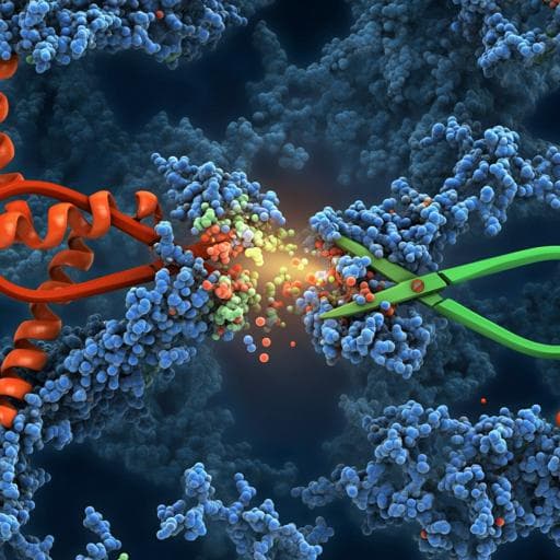
Characterising proteolysis during SARS-CoV-2 infection identifies viral cleavage sites and cellular targets with therapeutic potential
B. Meyer, J. Chiaravalli, et al.
This groundbreaking study by Bjoern Meyer and colleagues uncovers novel cleavage sites in SARS-CoV-2 proteins S and N using mass spectrometry. The research highlights potential therapeutic targets and demonstrates how targeting specific substrates inhibits SARS-CoV-2 replication. A must-listen for those interested in advancing COVID-19 research and treatments!
Related Publications
Explore these studies to deepen your understanding
Adjacent work that informs or extends this paper's methodology and findings.
Biology
CRISPR-Cas13d effectively targets SARS-CoV-2 variants, including Delta and Omicron, and inhibits viral infection
Z. Liu, X. Gao, et al.
Biology
Viral Population Heterogeneity and Fluctuating Mutational Pattern during a Persistent SARS-CoV-2 Infection in an Immunocompromised Patient
M. Brandolini, S. Zannoli, et al.
Biology
Protective roles and protective mechanisms of neutralizing antibodies against SARS-CoV-2 infection and their potential clinical implications
E. C. Abebe and T. A. Dejenie

Medicine and Health
A Young Female with SARS-CoV-2 Infection Presenting with Concurrent Neurological and Pulmonary Manifestations: A Case Report
P. Makuloluwa, K. P. Dissanayake, et al.


