Medicine and HealthFrontiers in Artificial Intelligence
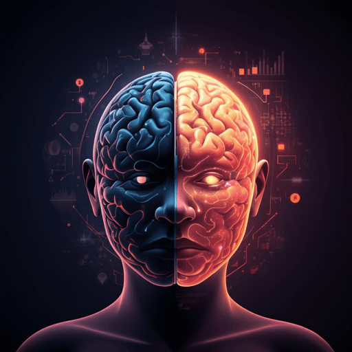
Can artificial intelligence improve the diagnosis and prognosis of disorders of consciousness? A scoping review
M. Bonanno, D. Cardile, et al.
AI (machine and deep learning) may transform how clinicians diagnose and prognosticate disorders of consciousness. This scoping review of 21 studies (14,683 patients, 180 controls) shows AI models using neurophysiology, neuroimaging, autonomic and clinical data to differentiate states (e.g., UWS vs MCS) and predict recovery, while calling for standardized data and demographic/etiological considerations. Research conducted by the authors listed in the <Authors> tag.
Related Publications
Explore these studies to deepen your understanding
Adjacent work that informs or extends this paper's methodology and findings.

Medicine and Health
The predictive performance of artificial intelligence on the outcome of stroke: a systematic review and meta-analysis
Y. Yang, L. Tang, et al.

Computer Science
Navigating the perils of artificial intelligence: a focused review on ChatGPT and responsible research and innovation
A. Polyportis and N. Pahos

Engineering and Technology
Artificial intelligence in the construction industry: A review of present status, opportunities and future challenges
S. O. Abioye, L. O. Oyedele, et al.
Medicine and Health
Artificial intelligence in mental health care: a systematic review of diagnosis, monitoring, and intervention applications
P. Cruz-gonzalez, A. W. He, et al.


