
Medicine and HealthJournal of Cerebral Blood Flow & Metabolism
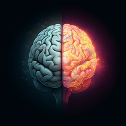
Brain water dynamics across sleep stages measured by near-infrared spectroscopy: Implications for glymphatic function
J. Yoon, M. Ji, et al.
Using near-infrared spectroscopy across the sleep–wake cycle, this study reports robust, state-dependent brain water fluctuations—rising from WAKE→NREM, falling from NREM→WAKE and NREM→REM, and rebounding at REM→NREM, with greater accumulation in early NREM cycles; research conducted by Authors present in <Authors> tag.
Related Publications
Explore these studies to deepen your understanding
Adjacent work that informs or extends this paper's methodology and findings.
Medicine and Health
A systematic review of alterations in sensorimotor networks following stroke: implications for integration and functional outcomes across recovery stages
N. S. A. Sahrizan, N. Yahya, et al.

Environmental Studies and Forestry
Competition for water induced by transnational land acquisitions for agriculture
D. D. Chiarelli, P. D'odorico, et al.

Environmental Studies and Forestry
A lighthouse to future opportunities for sustainable water provided by intelligent water hackathons in the Arabsphere
A. Batisha

Physics
Entangled photons enabled ultrafast stimulated Raman spectroscopy for molecular dynamics
J. J. Fan, Z. Ou, et al.


