
Medicine and HealthNature Communications
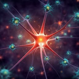
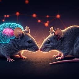
Brain mitochondrial diversity and network organization predict anxiety-like behavior in male mice
A. M. Rosenberg, M. Saggar, et al.
This groundbreaking research by Ayelet M. Rosenberg and colleagues explores how mitochondrial respiratory chain capacity influences stress-related behaviors in male mice. By analyzing 571 samples across 17 brain areas, the team identified critical mitochondrial networks linked to behavioral differences, unveiling distinct mitochondrial phenotypes relevant to behavior. Discover the intricate connections between mitochondria and behavior in the male mouse brain!
Related Publications
Explore these studies to deepen your understanding
Adjacent work that informs or extends this paper's methodology and findings.

Medicine and Health
Music prevents stress-induced depression and anxiety-like behavior in mice
Q. Fu, R. Qiu, et al.
Biology
Prefrontal Cortex-Specific Knockdown of Neurexin-1 in Rats Induces Anxiety-Like Behavior, Repetitive Behaviors, and Altered Social Interactions: A Proteomic Study
D. Wu, S. Zhang, et al.

Medicine and Health
Stress-induced vagal activity influences anxiety-relevant prefrontal and amygdala neuronal oscillations in male mice
T. Okonogi, N. Kuga, et al.
Psychology
Dynamic and stable hippocampal representations of social identity and reward expectation support associative social memory in male mice
E. Kong, K. Lee, et al.


