
BiologyNature Communications
Benchtop mesoSPIM: a next-generation open-source light-sheet microscope for cleared samples
N. Vladimirov, F. F. Voigt, et al.
Discover the groundbreaking mesoSPIM, a next-gen benchtop light-sheet microscope that enables high-resolution imaging of large cleared tissues. This innovative technology boasts enhanced field of view and versatility, making it valuable across fields like neuroscience and developmental biology, all crafted by a collaborative team of experts.
Related Publications
Explore these studies to deepen your understanding
Adjacent work that informs or extends this paper's methodology and findings.

Chemistry
Addressable nanoantennas with cleared hotspots for single-molecule detection on a portable smartphone microscope
K. Trofymchuk, V. Glembockyte, et al.
Biology
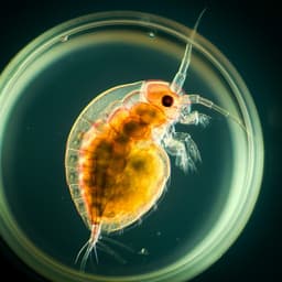
Zoobooth: A portable, open-source and affordable approach for repeated size measurements of live individual zooplankton
C. Broch and J. Heuschele

Engineering and Technology
A neuromorphic physiological signal processing system based on VO2 memristor for next-generation human-machine interface
R. Yuan, P. J. Tiw, et al.

Engineering and Technology
A neuromorphic physiological signal processing system based on VO₂ memristor for next-generation human-machine interface
R. Yuan, P. J. Tiw, et al.


