Medicine and Health
Attentional failures after sleep deprivation represent moments of cerebrospinal fluid flow
Z. Yang, S. D. Williams, et al.
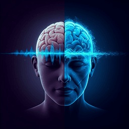
Sleep plays a fundamental role in maintaining brain health and cognitive performance, yet sleep deprivation is common and even a single night of lost sleep can cause marked cognitive impairment, including attentional failures where individuals fail to react to salient stimuli. Such lapses pose significant real-world risks (e.g., driving). Despite the reliable induction of attentional failures by sleep deprivation, the neural basis of these failures remains unclear. Prior work shows widespread neurophysiological effects of acute sleep deprivation: global BOLD fMRI fluctuations are altered and linked to electrophysiology, eyelid closures, and arousal state; lapses are associated with reduced activity in thalamus and cognitive control regions; and, locally, sleep-like low-frequency waves can occur in cortical patches in awake animals, with transient low-frequency increases predicting lapses in humans. These findings implicate brainwide hemodynamic changes and low-frequency neural oscillations in sleep-deprivation-induced attentional deficits, but the driver of spontaneous drops in arousal and behavior after sleep deprivation is unknown. One hypothesis is that lapses reflect brief engagement of a sleep-dependent function incompatible with waking behavior. Sleep supports clearance of neurotoxic waste products via CSF, with large low-frequency (0.01–0.1 Hz) CSF waves during NREM sleep. Sleep deprivation also modulates CSF pulsatility and solute concentrations, suggesting altered fluid dynamics after sleep loss, yet links between behavioral deficits and CSF flow have not been tested. To address this, the authors conducted a within-subject total sleep deprivation study in healthy adults using simultaneous fast fMRI, EEG, behavioral assessments (psychomotor vigilance test, PVT), and pupillometry to track neurophysiology and CSF dynamics. They discovered that, during wakefulness after sleep deprivation, sleep-like pulsatile CSF flow intrudes and couples with attentional deficits. Attentional failures coincide with a brain- and body-wide state change that underlies both behavioral deficits and pulsatile CSF flow, such that moments where attention fails signal a large-scale pulse of CSF flow in the brain.
The introduction synthesizes prior findings relevant to sleep deprivation and attention: (1) Sleep deprivation induces global BOLD fluctuations associated with changes in arousal and electrophysiology, including eyelid closures and vigilance fluctuations; (2) Attentional lapses are linked to reduced activation in thalamus and cognitive control networks; (3) Local sleep-like low-frequency waves occur in awake cortex (animals) and transient increases in low-frequency activity predict human lapses; (4) Sleep supports clearance of metabolic waste via CSF, which exhibits large low-frequency pulsations during NREM sleep; (5) Sleep deprivation affects CSF pulsatility and solute concentrations. However, these domains had not been integrated to test a direct link between attentional performance and CSF flow during wakefulness, particularly after sleep deprivation. Conflicting reports about clearance during sleep versus wake highlight differences in wake protocols (rested vs sleep-deprived) as a potential source of discrepancy, motivating the present investigation of CSF dynamics specifically under sleep deprivation and its behavioral consequences.
Design and participants: A within-subject study enrolled 26 healthy adults who completed two morning EEG–fast fMRI sessions (approximately 10 AM), one after a normal night of sleep at home (well-rested, WR) and one after a night of total sleep deprivation (SD) continuously supervised in the laboratory. The two sessions were 8–10 days apart. Overnight SD procedures included continuous monitoring with EEG and eye-tracking glasses, preventing prolonged eye closures (>2 s), and scheduled task blocks throughout the night. Inclusion criteria required regular sleep schedules and absence of sleep, medical, or psychiatric disorders; caffeine, alcohol, and sleep-aid abstention was enforced prior to scans. Written informed consent and IRB approval were obtained. Tasks and scanning protocol: Each visit included up to four 12-min PVT runs (auditory and visual variants, counterbalanced) followed by a 25-min eyes-closed resting-state run. PVT stimuli (0.5 s duration; auditory tone at 375 Hz or luminance-matched visual square replacing fixation) occurred with 5–10 s ISI; participants responded by button press. During resting-state, participants pressed a button on each breath as long as awake. Data acquisition: Imaging used a 3T Siemens Prisma with 64-channel head/neck coil. Anatomical scans were 1 mm isotropic multi-echo MPRAGE. Fast fMRI used single-shot gradient-echo multiband EPI (2.5 mm isotropic; TR=378 ms; MB=8; TE=31 ms; FA=37°; 40 interleaved slices; FOV=230×230; CAIPI FOV/4). Acquisition volumes were positioned to intersect the fourth ventricle, enabling measurement of ventricular CSF inflow alongside cortical BOLD. Systemic physiology was recorded (respiration belt, PPG) at 2000 Hz and synchronized to MRI. Pupillometry used an MR-compatible EyeLink 1000 at 1000 Hz; calibration was performed before each run. EEG was recorded with a 64-channel MR-compatible cap (BrainAmp MR) at 5000 Hz (scanner-synchronized), FCz reference, and complemented by a custom reference layer system for BCG artifact capture. EEG preprocessing and analysis: Gradient artifacts were removed via average artifact subtraction (moving average of 30 TRs), signals were common-average rereferenced (head and reference-layer electrodes separately), and BCG artifacts were regressed out using the reference-layer signals with sliding-window time-varying regression. Data were downsampled to 500 Hz, bandpass filtered (0.2–40 Hz), and ICA was used to remove eye/muscle components. Power spectral density (PSD) was computed via multitaper (Chronux) in 5 s windows with 5 tapers. Analyses emphasized POz due to consistent data quality. For spectrograms, power was normalized (z-scored within frequency). Oscillation-only spectrograms were derived by separating aperiodic and oscillatory components using FOOOF (1–25 Hz), with FDR-corrected significance testing. Sleep staging: Resting-state EEG was scored (AASM criteria) by two scorers using Visbrain; disagreements were resolved by a third scorer. Valid EEG was obtained in 24 subjects for sleep scoring and BOLD segmentation into stages (wake, N1, N2; N3 was rare and not analyzed due to only 5 subjects entering N3). MRI preprocessing: Cortical surfaces were reconstructed (FreeSurfer recon-all v6). Functional data were slice-time corrected (FSL) and motion-corrected to the middle frame (AFNI 3dvolreg). Runs were registered to anatomy (bbregister). Cortical gray matter ROIs were defined via FreeSurfer segmentation; time series were converted to percent signal change after discarding the first 20 volumes. CSF flow analysis: CSF inflow was measured from raw fMRI (slice-time corrected only; no motion correction to preserve inflow-sensitive slice position). Framewise displacement >0.5 mm segments were excluded. The fourth-ventricle CSF ROI was manually traced in the bottom three slices for each run. CSF signals were high-pass filtered >0.01 Hz and expressed as percent signal change relative to the 15th percentile value (to account for the inflow floor). CSF time series were upsampled to 4 Hz (spline interpolation) for event-locked averaging. Peak-locked and pupil coupling analyses: CSF peaks were identified in 0.01–0.1 Hz band-passed CSF as local maxima above 30% amplitude. Peak-locked epochs (±30 s) were extracted from z-scored, band-passed pupil traces. Significance was tested with time-binned two-sided t-tests versus baseline (−30 to −28 s), Bonferroni-corrected. Cross-correlations between pupil and CSF were computed and compared across conditions (WR vs SD; with vs without CSF peaks). Global cortical BOLD was examined for anticorrelation with pupil; an impulse response function (IRF) linking pupil to BOLD was estimated by convolving pupil traces with 1000 gamma-distribution IRFs and selecting the best correlation. Predicted CSF was generated by assuming pupil-locked BOLD fluctuations drive CSF (following prior work), without additional parameter fitting, and compared to measured CSF. Spectral power analysis: PSDs of BOLD and CSF were computed with multitaper (5 tapers) in 60 s segments classified by sleep stage. Low-frequency power (0.01–0.1 Hz) was compared across conditions using permutation tests across frequency bins (0.05 Hz bandwidth) with Bonferroni correction and paired t-tests for within-subject comparisons. Behavioral and omission-locked analyses: PVT performance (reaction times, omissions) from both auditory and visual tasks was analyzed. For behavior–CSF coupling, 60 s segments during confirmed wakefulness (EEG-verified; excluding eyelid closures >1 s and high motion) were categorized as high attention (all RT <500 ms), low attention (≥1 RT >500 ms), or omissions (≥1 miss). CSF low-frequency power was compared across categories (repeated-measures ANOVA; motion included as a factor). Omission-locked analyses (n=364 awake omissions, 26 subjects) excluded any trials during EEG-defined sleep or with eyelid closures >1 s. Event-locked EEG spectrograms (0.5–30 Hz), pupil, heart rate, respiratory rate/flow/volume, peripheral vascular volume, cortical BOLD, and CSF were computed relative to stimulus onset with baseline windows and Bonferroni/FDR-corrected statistics. To dissociate loss vs recovery of attention, omissions were classified: Type A (isolated; preceded and followed by valid responses), Type B (first of ≥3 consecutive omissions; onset of low attention), Type C (last of the series; recovery of attention). Eye-video verified wakefulness; analysis window −10 to +30 s, baseline −10 to −5 s. Systemic physiology: PPG and respiration were preprocessed (artifact spike removal; low-pass at 30 Hz and 10 Hz, respectively). Peripheral vascular volume (PVV) was computed as the standard deviation of PPG in 3 s windows. Heart rate was derived from interbeat intervals. Respiratory flow rate was computed from the derivative of the chest belt signal (rectified, low-pass 0.13 Hz); respiratory volume per time computed as the SD of respiration in 6 s windows. Bidirectional CSF flow replication experiment: An independent cohort (n=10; sleep-restricted to 4 h) underwent a single 10 AM scan with an acquisition tailored to measure both upward (inflow) and downward (outflow) CSF simultaneously (TR=593 ms; 2.5 mm; 8 slices; TE=31 ms; FA=51°; top slice at the narrow opening of the fourth ventricle; bottom slice at its base). ROIs were traced in the top 3 slices (outflow) and bottom 2 slices (inflow). Isolated omissions were used for event-locking. Outflow percent signal was inverted for display so that downward corresponds to downward flow in both traces.
- Sleep deprivation induces sleep-like low-frequency CSF pulsations during wakefulness: In resting-state wake segments (n=22 participants with ≥60 s wake), sleep-deprived wakefulness showed a 4.7 dB CSF power increase peaking at ~0.04 Hz, reaching magnitudes similar to N2 sleep (0.01–0.1 Hz power WR N2: −15.13 dB [95% CI −16.37, −13.88]; SD-wake: −14.96 dB [95% CI −15.88, −14.04]); paired analysis of wakefulness showed increased CSF low-frequency power after SD (n=18; p=0.0176). Cortical gray matter low-frequency BOLD power also increased after SD (p=0.0048, paired t-test). Motion did not differ across analyzed segments (p=0.43).
- Behavioral deficits with SD and CSF coupling: SD increased mean reaction time and the tail of RT distribution (more lapses; p<0.001, Mann–Whitney U) and increased omission rates (n=26; p<0.001, paired t-test). During PVT, higher CSF flow coincided with slower RTs and omissions. In 60 s wake segments categorized by performance, low-frequency CSF power was significantly higher in low-attention and omission segments than in high-attention segments (p<0.001 main effect; repeated-measures ANOVA; robust to motion).
- Pupil–CSF–behavior timing: Pupil diameter was significantly correlated with CSF (max r≈0.26 at lag −4.25 s; n=709 CSF peak segments), with constricted pupils linked to downward CSF signal and dilated pupils linked to subsequent upward CSF. Reaction times and omission rates increased before CSF peaks. Global cortical BOLD anticorrelated with pupil; the estimated pupil→BOLD IRF was negative with time-to-peak 6.75 s (consistent with vasoconstriction). Convolving pupil with the IRF significantly predicted CSF (zero-lag r=0.26; max r=0.30 at lag −1.75 s), supporting a vascular intermediary consistent with neuromodulatory state changes.
- Omission-locked biphasic CSF flow during wakefulness: Awake omission trials (n=364; 26 subjects) were locked to a downward CSF trough followed by an upward CSF peak. Gray matter BOLD showed a matched biphasic pattern. A replication with bidirectional CSF imaging (n=10; 114 isolated omissions) confirmed explicit CSF outflow beginning at the missed stimulus, followed by inflow at recovery.
- Electrophysiology during omissions: EEG spectrograms showed a broadband power drop around omissions, most pronounced in alpha–beta (10–25 Hz), followed by increases in slow wave activity (0.5–4 Hz) and alpha–beta power at recovery. Oscillation-only spectrograms retained significant clusters in slow and alpha–beta bands with widespread topography (centro-occipital predominance for alpha–beta; occipital/frontal for SWA).
- Systemic physiology coupling: Pupil constricted during omissions and dilated afterwards; heart rate and respiratory rate/flow/volume decreased at omission and increased at recovery; peripheral vascular volume showed biphasic changes. These brain–body signals were time-locked to omissions with corrected significance.
- Separating loss vs recovery of attention (Type A/B/C omissions): Type B (onset of series; arousal drop) was associated with decreased EEG alpha–beta power, reduced HR and respiratory rate, and CSF outflow (downward). Type C (end of series; arousal increase) showed increased broadband EEG power (including SWA during omission then alpha–beta rebound), increased HR and respiration, and CSF inflow (upward). Type A (isolated omission) showed a transient decrease then increase in EEG power and a biphasic CSF response aligned with behavioral recovery (~8–9 s later). Timing across modalities was consistent with vascular-mediated CSF dynamics following neuromodulatory shifts. Overall, attentional failures after SD are tightly coupled to large-scale, pulsatile CSF flow and coordinated neural and autonomic changes; attention drops when CSF flows outward and recovers when CSF flows inward.
The study directly links sleep-deprivation-induced attentional failures to pulsatile CSF dynamics during wakefulness. After SD, sleep-like CSF waves intrude into wake, reaching magnitudes typical of N1/N2 sleep, and these waves are tightly coupled to behavioral lapses, neural spectral shifts, and pupil constriction. The temporal ordering—pupil constriction and performance degradation preceding CSF peaks, with anticorrelated global BOLD—supports a mechanism where neuromodulatory state (likely including noradrenergic tone) modulates cerebrovascular dynamics that in turn drive CSF flow. Drops in ascending arousal could simultaneously suppress neuronal excitability (broadband EEG power reduction, alpha–beta suppression), constrict the pupil, alter systemic autonomic outputs (HR and respiration), and dilate cortical vessels, resulting in delayed downward CSF flow; attentional recovery reverses these processes, producing upward CSF flow. This integrated brain–body state model explains how brief attentional lapses after SD correspond to transient engagement of processes typically occurring in sleep (e.g., fluid transport). The results also have methodological implications for fMRI: regressing out CSF signals may inadvertently remove attention-related neural variance that is collinear with CSF dynamics, even in eyes-open wakefulness. The findings reconcile prior observations of altered global BOLD, low-frequency neural oscillations, and pupil-linked arousal with CSF transport, suggesting that destabilization of the ascending arousal system during SD leads to alternating epochs of reduced vigilance and enhanced fluid flow. Whether these state shifts serve functional clearance needs after prolonged wake (versus being epiphenomena of shared modulation) remains open, but the tight coupling and consistent timing align with a neuromodulatory vascular mechanism linking attention and CSF flow.
This work demonstrates that attentional failures after sleep deprivation are accompanied by large-scale, pulsatile CSF flow detectable in the fourth ventricle during wakefulness, together with coordinated changes in EEG spectral power, pupil diameter, and systemic physiology. Attention drops when CSF flows outward and recovers as CSF flows inward, consistent with neuromodulatory (likely noradrenergic) regulation of cerebrovascular dynamics driving CSF movement. The study identifies a central, integrated circuit linking attentional state and brain fluid dynamics and cautions that CSF regressors in fMRI can remove behaviorally relevant neural signals. Future directions include: causal tests of neuromodulatory contributions (e.g., pharmacology, LC-targeted interventions), higher-resolution measurements to localize cortical and perivascular dynamics, quantification of solute transport/clearance during omission-locked CSF pulses, assessment across diverse tasks and circadian phases, and evaluation of pupil-based proxies for noninvasive monitoring of CSF-related brain states.
- Initial fMRI protocol measured CSF inflow only; downward CSF flow could not be directly discriminated from reduced inflow in Experiment 1 (addressed by a separate bidirectional flow replication cohort).
- EEG and fMRI provide macroscopic measures; local sleep slow waves and precise circuit-level mechanisms could not be directly identified.
- Pupil diameter correlates with multiple neuromodulators, not solely noradrenergic tone; mechanistic attribution is indirect and based on timing and vascular signatures.
- Analyses are largely correlational and timing-based; causal inference about neuromodulation→vascular→CSF→behavior pathways is not established.
- N3 sleep was rare and not analyzed; generalization across deeper sleep stages not assessed.
- Some runs lacked valid eye-tracking; segments with motion or eyelid closures were excluded, potentially reducing data volume.
- The sample comprised healthy young adults; generalizability to other populations remains to be tested.
Related Publications
Explore these studies to deepen your understanding of the subject.