Medicine and HealthNature Communications
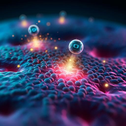
A region-confined PROTAC nanoplatform for spatiotemporally tunable protein degradation and enhanced cancer therapy
J. Gao, X. Jiang, et al.
This groundbreaking research by Jing Gao and colleagues introduces a novel PROTAC nanoplatform that overcomes traditional limitations in tumor specificity and pharmacokinetics. By integrating ROS-activatable and hypoxia-responsive components, this innovative strategy targets tumor cells effectively, improving drug release under specific conditions and leading to enhanced tumor eradication.
Related Publications
Explore these studies to deepen your understanding
Adjacent work that informs or extends this paper's methodology and findings.
Medicine and Health
CAR T-Cell Therapy for Cancer: Latest Updates and Challenges, with a Focus on B-Lymphoid Malignancies and Selected Solid Tumours
H. K. Tang, C. Tang, et al.
Medicine and Health
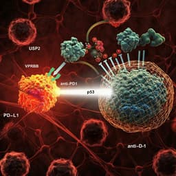
Targeting USP2 regulation of VPRBP-mediated degradation of p53 and PD-L1 for cancer therapy
J. Yi, O. Tavana, et al.
Medicine and Health
Advances in Photodynamic Therapy for the Treatment of Actinic Keratosis and Nonmelanoma Skin Cancer: A Narrative Review
A. S. Farberg, W. Justin, et al.
Biology
Tailored Functionalized Protein Nanocarriers for Cancer Therapy: Recent Developments and Prospects
R. J. Babu, A. K. Tiwari, et al.


