AgricultureScientific Reports
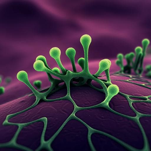
A rapid colorimetric LAMP assay for detection of *Rhizoctonia solani* AG-1 IA causing sheath blight of rice
P. Choudhary, P. Rai, et al.
Discover the rapid detection of *Rhizoctonia solani*, a major rice pathogen, through a groundbreaking colorimetric LAMP assay. This innovative method allows for confirmation of the pathogen in just 45 minutes, demonstrating high sensitivity and specificity. This research offers a crucial point-of-care diagnostic tool for timely intervention, conducted by Prassan Choudhary, Pallavi Rai, Jagriti Yadav, Shaloo Verma, Hillol Chakdar, Sanjay Kumar Goswami, Alok Kumar Srivastava, Prem Lal Kashyap, and Anil Kumar Saxena.
Related Publications
Explore these studies to deepen your understanding
Adjacent work that informs or extends this paper's methodology and findings.
Medicine and Health
A 3D-printed magnetic digital microfluidic diagnostic platform for rapid colorimetric sensing of carbapenemase-producing Enterobacteriaceae
P. Kanitthamniyom, P. Y. Hon, et al.

Medicine and Health
A nanoluciferase SARS-CoV-2 for rapid neutralization testing and screening of anti-infective drugs for COVID-19
X. Xie, A. E. Muruato, et al.

Physics
Ground-based and JWST Observations of SN 2022pul: II. Evidence from Nebular Spectroscopy for a Violent Merger in a Peculiar Type Ia Supernova
Kw, Jo, et al.

Agriculture
Exogenous metabolite application is a potential strategy for expanding the use of direct rice seeding with the aim of reducing seeding costs
B. Qing, Y. Jiang, et al.


