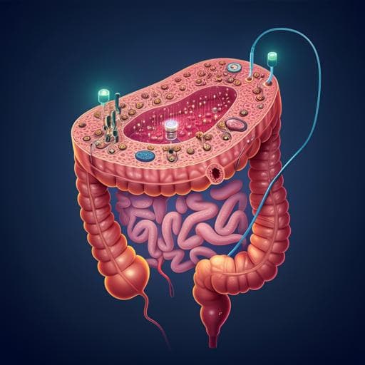
A microphysiological system for studying barrier health of live tissues in real time
R. Way, H. Templeton, et al.
Explore these studies to deepen your understanding
Adjacent work that informs or extends this paper's methodology and findings.
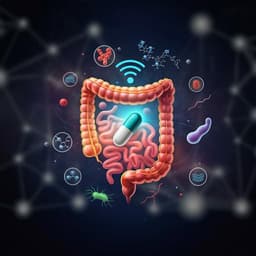
A self-powered ingestible wireless biosensing system for real-time in situ monitoring of gastrointestinal tract metabolites
E. D. L. Paz, N. H. Maganti, et al.
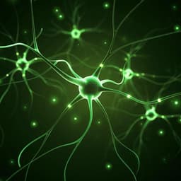
A fluorescent sensor for real-time measurement of extracellular oxytocin dynamics in the brain
D. Ino, Y. Tanaka, et al.

System characterization of a human-sized 3D real-time magnetic particle imaging scanner for cerebral applications
F. Thieben, F. Foerger, et al.

Development of prediction models for screening depression and anxiety using smartphone and wearable-based digital phenotyping: protocol for the Smartphone and Wearable Assessment for Real-Time Screening of Depression and Anxiety (SWARTS-DA) observational study in Korea
Y. Shin, A. Y. Kim, et al.


