Medicine and HealthNature Communications
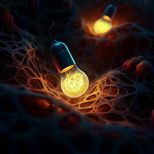
A biodegradable, flexible photonic patch for in vivo phototherapy
K. Deng, Y. Tang, et al.
Discover iCarP, a groundbreaking biodegradable photonic device that can illuminate internal tissues with unprecedented precision and adaptability! This innovative technology, developed by a team of talented researchers including Kaicheng Deng and Yao Tang, offers a novel solution for non-invasive phototherapies, promising safe and broad applications in medical diagnostics and treatment.
Related Publications
Explore these studies to deepen your understanding
Adjacent work that informs or extends this paper's methodology and findings.

Medicine and Health
In-vivo efficacy of biodegradable ultrahigh ductility Mg-Li-Zn alloy tracheal stents for pediatric airway obstruction
J. Wu, L. J. Mady, et al.
Biology
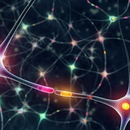
A genetically encoded tool for reconstituting synthetic modulatory neurotransmission and reconnect neural circuits in vivo
J. D. Hawk, E. M. Wisdom, et al.

Physics
Storage of photonic time-bin qubits for up to 20 ms in a rare-earth doped crystal
A. Ortu, A. Holzäpfel, et al.

Medicine and Health
Toward optical coherence tomography on a chip: in vivo three-dimensional human retinal imaging using photonic integrated circuit-based arrayed waveguide gratings
E. A. Rank, R. Sentosa, et al.


