Medicine and HealthNPG Asia Materials
2D carbon network arranged into high-order 3D nanotube arrays on a flexible microelectrode: integration into electrochemical microbiosensor devices for cancer detection
Y. Sun, X. Dong, et al.
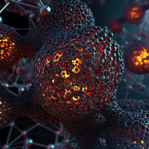
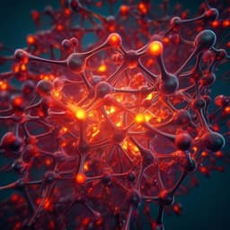
This groundbreaking research by Yimin Sun and colleagues showcases a novel mesoporous 2D carbon network engineered into 3D nanotube arrays, offering an innovative approach for high-performance electrochemical biosensing. This advancement not only enhances the detection of H₂O₂ from cancer cells but also enables real-time insights into cancer diagnostics and therapy efficacy.
Related Publications
Explore these studies to deepen your understanding
Adjacent work that informs or extends this paper's methodology and findings.
Medicine and Health
2D carbon network arranged into high-order 3D nanotube arrays on a flexible microelectrode: integration into electrochemical microbiosensor devices for cancer detection
Y. Sun, X. Dong, et al.

Engineering and Technology
A tactile sensor system with sensory neurons and a perceptual synaptic network based on semivolatile carbon nanotube transistors
S. Kim, Y. Lee, et al.
Engineering and Technology
Versatile self-assembled electrospun micro-pyramid arrays for high-performance on-skin devices with minimal sensory interference
J. Zhang, Z. Li, et al.
Engineering and Technology
Controlled on-chip fabrication of large-scale perovskite single crystal arrays for high-performance laser and photodetector integration
Z. Xu, X. Han, et al.


