
Medicine and HealthCell Death and Disease
USP18 is an essential regulator of muscle cell differentiation and maturation
C. S. Olie, A. Pinto-fernández, et al.
This groundbreaking study by Cyriel Sebastiaan Olie and colleagues uncovers the pivotal role of ubiquitin-specific protease 18 (USP18) in muscle cell differentiation. It reveals how USP18 influences both the initiation and maintenance of muscle cell formation, independent of its immune response role. Delve into the intricate relationship between gene expression and muscle physiology, as this research opens new avenues for understanding muscle pathologies.
Related Publications
Explore these studies to deepen your understanding
Adjacent work that informs or extends this paper's methodology and findings.

Medicine and Health
MOTS-c is an exercise-induced mitochondrial-encoded regulator of age-dependent physical decline and muscle homeostasis
J. C. Reynolds, R. W. Lai, et al.
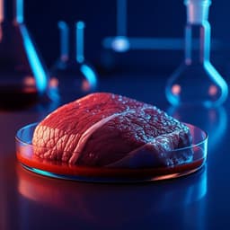
Food Science and Technology
Cell-based, cell-cultured, cell-cultivated, cultured, or cultivated. What is the best name for meat, poultry, and seafood made directly from the cells of animals?
W. K. Hallman, W. K. H. Ii, et al.

Education
Satisfaction as a key antecedent for word of mouth and an essential mediator for service quality and brand trust in international education
H. Stribbell and S. Duangekanong

Psychology
Is psilocybin an effective antidepressant and what is its Mechanism of action?
J. J. Mann


