Engineering and Technologynpj Microgravity
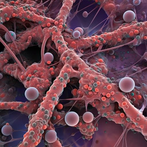
Unlocking the potential: analyzing 3D microstructure of small-scale cement samples from space using deep learning
V. Saseendran, N. Yamamoto, et al.
Discover groundbreaking research by Vishnu Saseendran, Namiko Yamamoto, Peter J. Collins, Aleksandra Radlińska, Sara Mueller, and Enrique M. Jackson on a novel deep learning method that reconstructs the 3D microstructure of cement samples studied in microgravity. The findings reveal unique characteristics of hydrated samples, with potential implications for material science and engineering.
Related Publications
Explore these studies to deepen your understanding
Adjacent work that informs or extends this paper's methodology and findings.
Medicine and Health
Design and Analysis of a Deep Learning Ensemble Framework Model for the Detection of COVID-19 and Pneumonia Using Large-Scale CT Scan and X-ray Image Datasets
X. Xue, S. Chinnaperumal, et al.

Medicine and Health
Recent Advancements and Perspectives in the Diagnosis of Skin Diseases Using Machine Learning and Deep Learning: A Review
J. Zhang, F. Zhong, et al.

Humanities
From remote sensing and machine learning to the history of the Silk Road: large scale material identification on wall paintings
S. Kogou, G. Shahtahmassebi, et al.

Medicine and Health
CovidCTNet: an open-source deep learning approach to diagnose covid-19 using small cohort of CT images
T. Javaheri, M. Homayounfar, et al.


