BiologyNature Communications
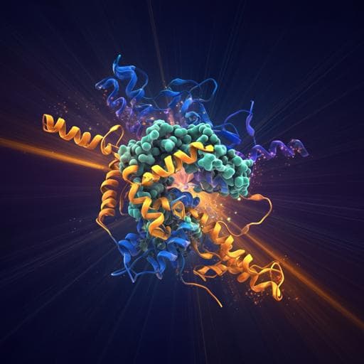
Ultraviolet optical horn antennas for label-free detection of single proteins
A. Barulin, P. Roy, et al.
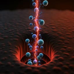
This groundbreaking research, conducted by Aleksandr Barulin, Prithu Roy, Jean-Benoît Claude, and Jérôme Wenger, explores a revolutionary nanophotonic platform that enables label-free detection of single proteins using their intrinsic UV autofluorescence. The study highlights the ability to monitor protein behaviors in real-time, paving the way for deeper insights into proteins in their native states.
Related Publications
Explore these studies to deepen your understanding
Adjacent work that informs or extends this paper's methodology and findings.
Engineering and Technology
Halftone spatial frequency domain imaging enables kilohertz high-speed label-free non-contact quantitative mapping of optical properties for strongly turbid media
Y. Zhao, B. Song, et al.
Medicine and Health
Optimized single-step optical clearing solution for 3D volume imaging of biological structures
K. Kim, M. Na, et al.
Medicine and Health
RapidET: a MEMS-based platform for label-free and rapid demarcation of tumors from normal breast biopsy tissues
A. V. G. K, G. Gogoi, et al.
Medicine and Health
Single-molecule amplification-free multiplexed detection of circulating microRNA cancer biomarkers from serum
S. Cai, T. Pataillot-meakin, et al.


