Medicine and HealthNature Medicine
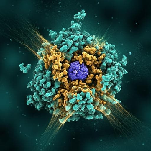
Trastuzumab deruxtecan in HER2-positive advanced gastric cancer: exploratory biomarker analysis of the randomized, phase 2 DESTINY-Gastric01 trial
K. Shitara, Y. Bang, et al.
Trastuzumab deruxtecan (T-DXd) has shown promising clinical advancements for patients with HER2+ gastric cancer, according to the DESTINY-Gastric01 trial. Engaging explorations into HER2 biomarkers and resistance mechanisms highlight correlations that could shape future therapies. This vital research was led by an exceptional team of authors.
Related Publications
Explore these studies to deepen your understanding
Adjacent work that informs or extends this paper's methodology and findings.
Medicine and Health
Trastuzumab deruxtecan versus trastuzumab emtansine in HER2-positive metastatic breast cancer: long-term survival analysis of the DESTINY-Breast03 trial
J. Cortés, S. A. Hurvitz, et al.

Medicine and Health
Trastuzumab deruxtecan in HER2-positive advanced breast cancer with or without brain metastases: a phase 3b/4 trial
N. Harbeck, E. Ciruelos, et al.

Medicine and Health
Biomarker-directed targeted therapy plus durvalumab in advanced non-small-cell lung cancer: a phase 2 umbrella trial
B. Besse, E. Pons-tostivint, et al.

Medicine and Health
Entrectinib in ROS1-positive advanced non-small cell lung cancer: the phase 2/3 BFAST trial
S. Peters, S. M. Gadgeel, et al.


