Medicine and Health
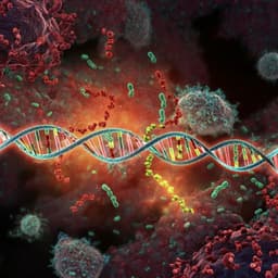
Transplacental transmission of SARS-CoV-2 infection
A. J. Vivanti, C. Vauloup-fellous, et al.
SARS-CoV-2 causes COVID-19 and is mainly transmitted through respiratory droplets, though other routes have been hypothesized. Reports of perinatal transmission exist, but the exact transmission route (transplacental versus transcervical or environmental) has been unclear. Establishing whether and how SARS-CoV-2 reaches the fetus is important to prevent neonatal infection, guide pregnancy management, and understand viral biology. This paper presents a comprehensive case study demonstrating transplacental transmission of SARS-CoV-2 with neonatal clinical manifestations consistent with COVID-19-related neurological signs and symptoms.
Prior reports described possible perinatal transmission but often lacked systematic testing of placenta, amniotic fluid, and maternal/newborn blood, or only reported neonatal antibodies without viral detection. Using a proposed classification system for SARS-CoV-2 infection in pregnancy, congenital infection is proven if virus is detected in amniotic fluid collected before membrane rupture or in early neonatal blood. Many earlier cases would thus be only possible or unlikely. Similar placental findings have been observed in SARS-CoV-1 (intervillositis and fibrin deposition). ACE2, the receptor for SARS-CoV-2, is expressed in placental tissues and may peak around late gestation to early postnatal life, supporting biological plausibility of transplacental transmission in late pregnancy.
Design: Single comprehensive case study of a pregnant woman with COVID-19 and her neonate, including clinical, virological, imaging, and pathological investigations. Patient sampling: Maternal nasopharyngeal and vaginal swabs obtained per CDC guidance; clear amniotic fluid collected prior to rupture of membranes during cesarean section; placental tissue sampled from the chorionic side and homogenized; maternal and neonatal blood collected; neonatal nasopharyngeal and rectal swabs collected at 1 hour of life (after cleaning), and repeated at days 3 and 18; non-bronchoscopic bronchoalveolar lavage (BAL) performed using standardized pediatric technique while intubated, with 1 mL/kg warmed saline instilled and aspirated from each lung without ventilator disconnection. Specimen handling: Blood and swabs placed in Virocult viral transport media; all specimens stored at +4 °C and tested within 24 hours. RT-PCR: Viral RNA extracted from 200 µL of clinical samples using NucliSENS easyMag, eluted in 100 µL. RealStar SARS-CoV-2 RT-PCR Kit 1.0 (Altona Diagnostics) targeting E gene (lineage B betacoronavirus) and S gene (SARS-CoV-2 specific) used with internal positive control. Thermal cycling: 55 °C 20 min (RT), 95 °C 2 min, then 45 cycles of 95 °C 15 s, 55 °C 45 s, 72 °C 15 s on Applied Biosystems ViiA7. Positivity defined as Ct < 40. Limit of detection ~1200 copies/mL (12 copies/reaction). Assay reproducibility and inter-assay agreement 100% vs two other techniques. Positive control: SARS-CoV-2 culture supernatant. Placental pathology: Gross and microscopic examination per Amsterdam Consensus. Placenta fixed in 4% buffered formalin, paraffin-embedded; sections 3–5 µm stained with HES, PAS, and Gomori-Grocott. Immunohistochemistry (Leica Bond III) with peroxidase detection and hematoxylin counterstain; antigens included CD68, CD163, CD20, CD3, CD5, CMV, Parvovirus, and SARS-CoV-2 (anti-N protein). Negative controls included omission of primary antibody and SARS-CoV-2 negative placentas with similar fixation. Clinical neonatal assessments: Immediate resuscitation per international guidelines; NICU isolation in negative pressure room; monitoring with ECG, end-tidal CO2, SpO2, perfusion index; Sarnat scoring, point-of-care echocardiography, lung ultrasound; serial blood gases and routine labs; CSF analysis and cultures when neurological symptoms developed; EEG and cerebral ultrasound; brain MRI at 11 days of life; follow-up clinical and MRI evaluation at ~2 months. Ethics: Written informed consent obtained. According to French regulations, IRB approval not required for case reports with consent; reviewed by French Ethical Committee for Obstetrics and Gynecology.
- Maternal infection: A 23-year-old at 35+2 weeks presented with fever and respiratory symptoms. RT-PCR detected SARS-CoV-2 E and S genes in maternal blood, nasopharyngeal, and vaginal swabs. Maternal labs showed thrombocytopenia (54 × 10^9/L), lymphopenia (0.54 × 10^9/L), prolonged APTT (60 s), elevated AST (81 IU/L), ALT (41 IU/L), CRP (37 mg/L), and ferritin (431 µg/L).
- Delivery: Category III fetal heart rate tracing prompted category II cesarean section at 35+5 weeks under isolation. Amniotic fluid collected before membrane rupture tested positive for SARS-CoV-2 (E and S genes).
- Neonatal virology: Early neonatal blood and non-bronchoscopic BAL fluid positive for SARS-CoV-2 (E and S genes). Serial nasopharyngeal and rectal swabs at 1 h, day 3, and day 18 were all positive. Blood cultures negative.
- Viral loads (Log copies): Placenta 11.15 (copies/million cells); maternal: blood 4.87, nasopharyngeal 4.22, vaginal 0.63, amniotic fluid 2.09. Neonatal: blood 1.15; nasopharyngeal swab day 1: 2.21, day 3: 7.30, day 18: 4.54; rectal swab 4.71. Placental viral load was markedly higher than other compartments.
- Placental pathology: Diffuse perivillous fibrin deposition with infarction and acute and chronic intervillositis. Immunohistochemistry showed intense cytoplasmic positivity for SARS-CoV-2 N-protein in perivillous trophoblastic cells. No other pathogens detected.
- Neonatal clinical course: Male late-preterm (2540 g) required brief ventilation; extubated at ~6 h. Vital signs stable; early echocardiography, lung ultrasound, and Sarnat score normal. On day 3, sudden irritability, poor feeding, axial hypertonia, opisthotonos. First CSF: negative PCR for SARS-CoV-2 and other pathogens; pleocytosis (300 leukocytes/mm^3) and mildly elevated protein (1.49 g/L) with normal glucose; blood culture sterile. EEG and cerebral ultrasound normal. Symptoms improved over 3 days; second CSF (day 5) normal; mild hypotonia and feeding difficulty persisted.
- Neuroimaging: Brain MRI at day 11 showed bilateral periventricular and subcortical white matter gliosis (slight left predominance). Follow-up at ~2 months showed improved neurological exam and reduced white matter injury on MRI.
- Feeding: Formula only (no breastfeeding).
- Overall: Evidence fulfilled criteria for proven congenital (transplacental) SARS-CoV-2 infection: virus detected in amniotic fluid collected prior to membrane rupture and in early neonatal blood; high placental viral load with inflammatory pathology; subsequent neonatal viremia and clinical neurological manifestations.
This case demonstrates proven congenital SARS-CoV-2 infection via the transplacental route. Detection of viral RNA in amniotic fluid collected before membrane rupture, high viral load and antigen in placental tissue with intervillositis, and early neonatal blood positivity together indicate maternal viremia leading to placental infection and neonatal viremia. Rising neonatal nasopharyngeal viral loads between day 1 and day 3 while in strict isolation further support true neonatal infection rather than contamination. Findings align with prior observations of placental inflammation in SARS-CoV-1 and are biologically plausible given ACE2 expression in placental tissues, which may peak around late gestation. Clinically, the neonate developed neurological symptoms with CSF inflammation and white matter injury on MRI, consistent with neuroinflammatory/vasculitic processes described in adult COVID-19. Compared with earlier reports where sampling was incomplete, this case meets proposed classification criteria for proven congenital infection, strengthening evidence that transplacental transmission can occur in late pregnancy and can be associated with neonatal neurological involvement.
Transplacental transmission of SARS-CoV-2 is possible in the last weeks of pregnancy and may lead to placental inflammation and neonatal viremia. Neonatal neurological symptoms, likely related to cerebral vasculitis or inflammatory injury, may occur. Comprehensive maternal, placental, and neonatal testing should be performed in suspected cases. Future research should define the incidence, risk factors, timing across gestation, mechanisms of placental infection and inflammation, and long-term neurodevelopmental outcomes in exposed infants.
Single-case report limits generalizability and precludes incidence estimation. While findings strongly support transplacental transmission in late pregnancy, the study cannot determine the risk or consequences of infection earlier in gestation. Some neonatal samples (e.g., CSF) were negative by PCR despite clinical signs, which may reflect compartmentalization or timing of sampling. Broader cohorts with standardized, systematic sampling are needed.
Related Publications
Explore these studies to deepen your understanding of the subject.

