
BiologyAccounts of Chemical Research
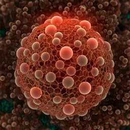
Toward Compositional Contrast by Cryo-STEM
M. Elbaum, S. Seifer, et al.
Discover the transformative potential of scanning transmission electron microscopy (STEM) for cryo-electron microscopy in biological research, as explored by authors Michael Elbaum, Shahar Seifer, Lothar Houben, Sharon G. Wolf, and Peter Rez. This innovative approach not only preserves native elemental composition but also enhances compositional contrast, revealing new insights in the life sciences.
Related Publications
Explore these studies to deepen your understanding
Adjacent work that informs or extends this paper's methodology and findings.

Biology
Toward Compositional Contrast by Cryo-STEM
M. Elbaum, S. Seifer, et al.
Medicine and Health
An instantly fixable and self-adaptive scaffold for skull regeneration by autologous stem cell recruitment and angiogenesis
G. Lu, Y. Xu, et al.

Earth Sciences
Enhanced Arctic moisture transport toward Siberia in autumn revealed by tagged moisture transport model experiment
T. Sato, T. Nakamura, et al.
Biology
Single-particle cryo-EM structures from IDPC-STEM at near-atomic resolution
I. Lazić, M. Wirix, et al.


