Medicine and HealthNature Communications
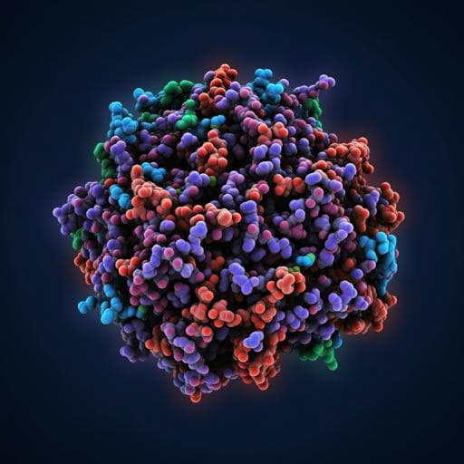
Structural plasticity of SARS-CoV-2 3CL Mpro active site cavity revealed by room temperature X-ray crystallography
D. W. Kneller, G. Phillips, et al.
Dive into the exciting findings of groundbreaking research conducted by Daniel W. Kneller and colleagues that reveals the room-temperature X-ray structure of unliganded SARS-CoV-2 3CL Mpro. This study highlights the significant implications for antiviral inhibitor development and enhances our understanding of viral replication dynamics.
Related Publications
Explore these studies to deepen your understanding
Adjacent work that informs or extends this paper's methodology and findings.

Medicine and Health
Identification of SARS-CoV-2 Mpro inhibitors containing P1' 4-fluorobenzothiazole moiety highly active against SARS-CoV-2
N. Higashi-kuwata, K. Tsuji, et al.

Chemistry
A sustainable synthesis of the SARS-CoV-2 Mpro inhibitor nirmatrelvir, the active ingredient in Paxlovid
J. R. A. Kincaid, J. C. Caravez, et al.
Medicine and Health
Histones released by NETosis enhance the infectivity of SARS-CoV-2 by bridging the spike protein subunit 2 and sialic acid on host cells
W. Hong, J. Yang, et al.
Medicine and Health
Broad ultra-potent neutralization of SARS-CoV-2 variants by monoclonal antibodies specific to the tip of RBD
H. Ma, Y. Guo, et al.


