Medicine and HealthCommunications Chemistry
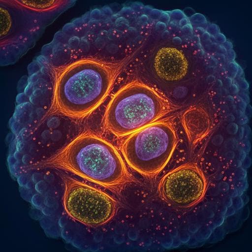
Specific intracellular signature of SARS-CoV-2 infection using confocal Raman microscopy
H. Salehi, A. Ramoji, et al.
Discover groundbreaking research by Hamideh Salehi and colleagues as they unveil a virus-specific Raman molecular signature for SARS-CoV-2, differentiating infected from non-infected cells with remarkable accuracy. This study highlights increased tryptophan levels as a potential biomarker for COVID-19, showcasing the transformative power of Raman spectroscopy in virus-host studies.
Related Publications
Explore these studies to deepen your understanding
Adjacent work that informs or extends this paper's methodology and findings.

Medicine and Health
Lethality of SARS-CoV-2 infection in K18 human angiotensin-converting enzyme 2 transgenic mice
F. S. Oladunni, J. Park, et al.
Medicine and Health
Systematic detection of co-infection and intra-host recombination in more than 2 million global SARS-CoV-2 samples
O. A. Pipek, A. Medgyes-horváth, et al.
Medicine and Health
Broad ultra-potent neutralization of SARS-CoV-2 variants by monoclonal antibodies specific to the tip of RBD
H. Ma, Y. Guo, et al.
Biology
The Breadth of the Neutralizing Antibody Response to Original SARS-CoV-2 Infection is Linked to the Presence of Long COVID Symptoms
A. M. Buck, A. N. Deitchman, et al.


