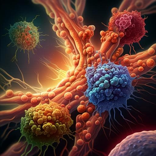
Single-cell and spatial transcriptome analyses reveal tertiary lymphoid structures linked to tumour progression and immunotherapy response in nasopharyngeal carcinoma
Y. Liu, S. Ye, et al.
Explore these studies to deepen your understanding
Adjacent work that informs or extends this paper's methodology and findings.

Single cell analysis reveals distinct immune landscapes in transplant and primary sarcomas that determine response or resistance to immunotherapy
A. J. Wisdom, Y. M. Mowery, et al.
Prolonged response to first-generation tyrosine kinase inhibitor in a metastatic non-small cell lung cancer harbouring complex G719X and S768I mutations: A case report from Vietnam and literature review
K. H. Do, D. T. Le, et al.
CD317 maintains proteostasis and cell survival in response to proteasome inhibitors by targeting calnexin for RACK1-mediated autophagic degradation
J. Cheng, G. Zhang, et al.

Integrating digital pathology and mathematical modelling to predict spatial biomarker dynamics in cancer immunotherapy
L. G. Hutchinson and O. Grimm


