Medicine and HealthCell Death Discovery
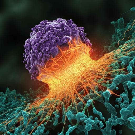
ROS induces NETosis by oxidizing DNA and initiating DNA repair
D. Azzouz, M. A. Khan, et al.
Discover how reactive oxygen species play a crucial role in the formation of neutrophil extracellular traps! This research, conducted by Dhia Azzouz, Meraj A. Khan, and Nades Palaniyar, dives into the mechanisms of NETosis, revealing how DNA damage and ROS lead to this fascinating phenomenon.
Related Publications
Explore these studies to deepen your understanding
Adjacent work that informs or extends this paper's methodology and findings.
Medicine and Health
ROS induces NETosis by oxidizing DNA and initiating DNA repair
D. Azzouz, M. A. Khan, et al.
Medicine and Health
SK3 channel and mitochondrial ROS mediate NADPH oxidase-independent NETosis induced by calcium influx
D. N. Douda, M. A. Khan, et al.
Medicine and Health
Histones released by NETosis enhance the infectivity of SARS-CoV-2 by bridging the spike protein subunit 2 and sialic acid on host cells
W. Hong, J. Yang, et al.
Medicine and Health
Failure of DNA double-strand break repair by tau mediates Alzheimer's disease pathology in vitro
M. Asada-utsugi, K. Uemura, et al.


