Food Science and Technology
Reprocessing seafood waste: challenge to develop aquatic clean meat from fish cells
Y. Tsuruwaka and E. Shimada
In recent years, the technology of 3D cell culture has been developed and applied to a cell sheet. Although these technologies have been applied in the medical field, they are expected to be applied in other fields, such as the food industry. Our aim is to explore applying this technology to produce marine food resources. As we currently face on the food crisis, population growth is resulting in a rapidly increasing demand for livestock products, which leads to environmental stress since raising livestock requires large amounts of natural resources and accounts for about 14.5% of total anthropogenic greenhouse gas emissions. Therefore, the establishment of a sustainable food resource production system for the next generation will be beneficial for environmental and ecological protection. In addition, a sustainable food production system is expected to reduce waste by recycling it effectively.
Fish is increasingly consumed worldwide, resulting in enormous impacts on the ecosystem, including overfishing. We have reported the farming of edible deep-sea fish that are difficult to rear, and their biological activity at the cellular level. Fish farming needs to take into account its operating costs and rearing space. In the present study, we focused on the recycling of fish waste material and its possibility to develop ‘aquatic clean meat’ from fish cells to meet the challenge outlined in the United Nations’ (UN) Sustainable Development Goal 14: “Conserve and sustainably use the oceans, seas and marine resources for sustainable development”. We focused on fish fins because 1) they are often discarded as food waste and 2) collecting fresh fins partially does not unnecessarily take the lives of fish. If the technology were developed to create ‘aquatic clean meat’ from seafood waste, it would be an environmentally friendly way to increase animal welfare and sustainability. As a first step, we demonstrated the potential for and the advantages of obtaining cells from fins at the individual level here.
Fish have a high level of ability to regenerate. They are able to regenerate various body parts, including fins, which are equivalent to the hand and foot in humans, as well as hearts, neurons, and so on. In general, regeneration occurs by ‘dedifferentiation’ and/or self-renewal with basal stem cells. Individual fish are able to regenerate their partially lost fins within a few weeks. Thus, fish can be a highly regenerative food material. In this study, we deal with partial fins cut from a fish’s body. Cultured fin tissues cut from fish bodies are known to show explants around the tissues in a culture flask. Therefore, we investigated the cell variations that have experienced the regeneration process, including the differentiation potential if the cells have dedifferentiated.
In general, when embryonic stem (ES) cells (i.e., the inner cell mass of early embryos as well as amphibian undifferentiated embryonic cells) are cultured for differentiation induction in vitro, they are known to differentiate neural cells unless cultured with specific stimulating factors, that is, external signals such as serum, growth factors, etc. Thus, the basal state of undifferentiated cells was destined to neural induction intrinsically. Shimada et al. reported that fins were derived from the somatic mesoderm which developed from somatic stem cells. This suggests the possibility that when fin cells in the regeneration process are dedifferentiated and become undifferentiated cells, they may become basal neural somatic stem cells. If dedifferentiated fish cells have the ability to differentiate to various types of cells, such as muscle, fat, fibers, and nerves, which are the main components of edible animal meat, then we can manipulate and arrange the proportion of these meat components to create ‘aquatic clean meat’. We obtained cells from the fins of waste materials and investigated their potential for cell differentiation, which could be applied to clean meat.
The authors situate their work within several bodies of literature: (1) advances in 3D cell culture and cell-sheet-based tissue engineering with applications in medicine that could translate to food production; (2) environmental impacts of conventional livestock production and the potential of cultured meat to mitigate greenhouse gas emissions; (3) overfishing and the need for sustainable marine resource use; (4) fish regenerative biology, including fin, heart, and neuronal regeneration via dedifferentiation and stem cell activity; (5) the intrinsic tendency of ES cells to adopt neural fates in the absence of external inhibitors, motivating exploration of neural differentiation potential in dedifferentiated fin-derived cells; and (6) prior establishment of fish fin-derived cell lines and observations of explant outgrowth, as well as broader discussions on the promise and challenges of cellular agriculture and cell-based seafood. These references frame the hypothesis that fin-derived cells, potentially exhibiting dedifferentiation, might be coaxed into multiple edible meat components via simple culture manipulations for sustainable food production.
Animal: Live thread-sail filefish Stephanolepis cirrhifer was captured near Jogashima Island in Yokosuka City, Kanagawa Prefecture, Japan. Specimens were maintained in aerated artificial seawater at 34.0 ppt salinity and 20 ± 2 °C. All procedures were approved by the Animal Experimental Committee of JAMSTEC and followed animal care guidelines.
Preparation of the cell line: A 5 mm² fin tissue piece was obtained and placed in 70% ethanol, washed three times with PBS, then six times with penicillin/streptomycin on ice. The fin tissue was cut into ~1 mm squares, incubated in 0.25% Trypsin–0.02% EDTA for 20 min at room temperature, centrifuged at 1100 rpm, and washed twice with Leibovitz's L-15 medium. Tissue was immersed in L-15 with 10% fetal bovine serum (FBS) and 1% Zell Shield, seeded in a 25 cm² Collagen I Coated flask, and cultured at 25 °C. After 24 h, outgrowth around tissue was confirmed. Several days later, cells were detached with TrypLE Express and reseeded. Media was replaced every 3 days and cells were subcultured at 4.0 × 10^5 cells/ml for five passages to establish the line, named deSC cells. The line was deposited at NITE (accession NITE BP-1369). Morphology was observed with an Olympus CKX41 inverted microscope and ARTCAM-300MI-WOM camera.
Induction of differentiation: 4.0 × 10^5 deSC cells were seeded in L-15 with 10% FBS in 25 cm² Collagen I Coated or non-coated flasks. After 24 h, cells were washed with PBS and treated as follows: Collagen I Coated flasks received AIM V + 10% FBS, L-15 + 10% heat-inactivated SeaGrow (salmon serum), or KBM Neural Stem Cell medium + 1X Neural Induction Supplement. Non-coated flasks received L-15 only. Differentiation was imaged every 60 s to generate time-lapse movies (Axio Vision v4.8).
Lipid staining: Cells were fixed in 4% paraformaldehyde in PBS at room temperature for 1 h. BODIPY staining (Adipocyte Fluorescent Staining kit) was performed per manufacturer’s protocol. For Oil Red O, fixed cells were treated with 60% isopropanol for 5 min, incubated with Oil Red O for 25 min at room temperature, and imaged.
Immunofluorescence: Differentiated cells were fixed in 4% paraformaldehyde in PBS at room temperature for 12 h, incubated with Milli-Mark FluoroPan Neuronal Marker (Mouse IgG-Alexa 488) per instructions, and imaged on a Zeiss Axio Observer.D1 with AxioCam HRc; images analyzed in AxioVision v4.8.
Scanning electron microscopy (SEM): Adherent cells on Collagen I Coated slides were fixed overnight at 4 °C in 2.5% glutaraldehyde in culture medium, postfixed in 2.0% osmium tetroxide in PBS for 2 h at 4 °C, dehydrated through graded ethanol, freeze dried (VFD-21S), coated with osmium (POC-3 osmium plasma coater), and observed with a JSM-6700F field emission SEM at 5 kV.
Gas chromatography: Fatty acids in fin, fin cells, and differentiated cells were analyzed by injecting 1 µl extract into an Agilent 7890A GC equipped with an Omega wax 320 30 m × 0.32 mm column.
Culture conditions overview (examples used in results): Cells were cultured at 25 °C without CO2. Example combinations: Fibroblast-like (L-15 + FBS, Collagen I Coated); Skeletal muscle-like (AIM V + FBS, Collagen I Coated); Neural-like (L-15 without serum, non-coated); Neurofilament (KBM + Neural Induction Supplement, Collagen I Coated); Adipocyte (L-15 + SeaGrow, Collagen I Coated); Spheroid (L-15 + FBS, spheroid cultureware); CoCoon (L-15 + horse serum, non-coated).
- Fin-derived fibroblast-like cells from Stephanolepis cirrhifer (deSC) were established from fin explants; stable fibroblast-like cells obtained by passage 5 and maintained up to 350 passages at 25 °C without CO2, indicating immortalization.
- Chromosomal analysis (Q-banding) showed 33 chromosomes (2n = 30 + X,X,Y) in 96% of deSC cells, matching wild type S. cirrhifer; 4% showed 66 chromosomes (likely cells in division with replicated DNA).
- Differentiation depended on simple culture parameters (medium, serum, ECM), without genetic manipulation: cells adopted skeletal muscle-like, neural-like, neurofilament, adipocyte, spheroid, and CoCoon morphologies under distinct conditions.
- In L-15 medium without serum on non-coated flasks, deSC cells differentiated into neural-like cells within 24 h, consistent with neural default in absence of inhibitors.
- Direct neural induction using KBM Neural Stem Cell medium + Neural Induction Supplement produced neurofilaments with maximum length 465 µm and average elongation speed 45.71 µm/h; neural immunofluorescence supported neuronal identity.
- SeaGrow (salmon serum) induced adipocyte differentiation: cells became round with intracellular white droplets (0.5–2.0 µm), confirmed as lipid by Oil Red O, BODIPY staining, and gas chromatography.
- 3D culture: deSC cells formed spheroids (20–300 µm) by incorporating surrounding cells; with horse serum, formed adherent migratory aggregates termed “CoCoon,” moving at 38.46 µm/h and undergoing major movement/fusion approximately every 3.5 h; CoCoon size ranged 20–1000 µm. Spheroids and CoCoon were stable for at least three weeks.
- Differentiations (spheroids, CoCoon, skeletal muscle-like, adipocytes) were reversible to fibroblast-like state upon returning to basic culture (L-15 + 10% FBS, Collagen I Coated); neural differentiation was not reversible under tested conditions.
- Multi-layered cell-sheet cultures were produced from deSC cells, including adipocyte cell sheets under adipogenic conditions; sheets could be detached and shaped.
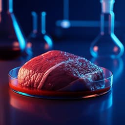
- Prototype “aquatic clean meat” (sashimi-like) was fabricated from stacked deSC cell sheets approximately 70 mm × 30 mm × 2 mm. Simple sensory assessment: white color, no smell, no taste, smooth texture, soft firmness; shape/size flexible but still distinct from real sashimi.
- deSC cells expressed markers associated with pluripotency (AP, Nanog, Oct4, SSEA-3, TRA-1-60) in supplementary data, suggesting inherent pluripotent-like properties.
- Similar 2D differentiation phenomena observed in 57 fish cell types (including Sebastiscus marmoratus), leading authors to term such cells “iPX cells” (inherently Pluripotent X cells).
- Cell line deposited at NITE (NITE BP-1369).
The study addresses the challenge of creating sustainable, animal-welfare-friendly seafood by demonstrating that fin tissues—typically discarded as waste—yield cells capable of extensive proliferation and differentiation into multiple meat-relevant lineages through simple culture manipulations. By showing that medium composition, serum type, and ECM alone can drive deSC cells into neural-like, muscle-like, adipogenic, and organized 3D aggregates, the authors provide a minimal, scalable toolkit for designing cell compositions and structures relevant to food texture and function. The capacity to form multilayered sheets and 3D constructs (spheroids, CoCoon) and to reversibly toggle many differentiated states supports practical processing and shaping workflows for cultivated seafood products. Neural differentiation following neural-default logic and direct induction underscores possible intrinsic pluripotent-like plasticity in fish fin-derived cells, potentially simplifying processes compared to mammalian systems that often require genetic reprogramming. The prototype sashimi demonstrates feasibility of assembling macro-scale edible constructs from ~20 µm cells, with basic sensory attributes recorded. The authors argue that this simplicity, combined with easy-to-source raw materials (fins/skin), controlled clean production (reduced microplastic contamination risk), and suitability for limited-space environments, makes aquatic clean meat a promising path to sustainable seafood, while also offering biological insight into fish diversity and regenerative capacity.
This work establishes fin-derived deSC cells from Stephanolepis cirrhifer as a versatile, robust platform for cultivated seafood: the cells proliferate long-term without CO2, maintain karyotypic integrity, and undergo multiple differentiations and 3D organizations via simple, non-genetic stimuli. Leveraging these properties, the authors fabricated a prototype sashimi-like construct from stacked cell sheets, illustrating a pathway to reprocess seafood waste into value-added, sustainable food. Future research should focus on detailed identification and characterization of differentiated lineages and sheet-assembled tissue components (e.g., muscle, adipose, neurofilaments, muscle proteins, collagen), optimization of cell-type ratios to tailor taste and texture to consumer preferences, improving resemblance to real sashimi, refining processing and scalability, and expanding species coverage to harness broadly available iPX-like fish cells for consistent, safe, and sustainable production.
- The study does not deeply identify or characterize differentiated cell types beyond morphology and selected stains/markers; authors note that the work does not focus on cell identification and that further examination is required.
- The sashimi-like prototype is described as still very different from real sashimi; sensory evaluation was informal and limited.
- While differentiation reversibility was shown for several lineages, neural differentiation was not reversible under reported conditions, and functional maturity of differentiated cells was not assessed.
- Serum screening was limited; some mammalian sera (rabbit, sheep) did not support stable outcomes (data not shown), and broader serum-free/defined conditions were not explored in detail.
- Food safety, scalability, cost analysis, and long-term production stability were not evaluated in this study.
Related Publications
Explore these studies to deepen your understanding of the subject.