Medicine and Health
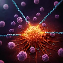
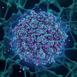
Reformulating lipid nanoparticles for organ-targeted mRNA accumulation and translation
K. Su, L. Shi, et al.
The study addresses a central challenge in mRNA therapeutics: achieving true organ targeting that requires both selective accumulation of mRNA-loaded nanoparticles and efficient translation within the target organ while minimizing off-target exposure, especially in the liver. Conventional lipid nanoparticles (LNPs) show strong hepatic tropism due to their canonical four-component composition (ionizable lipid, phospholipid, cholesterol, PEG-lipid), which contributes to similar physicochemical properties across systems and persistent liver accumulation after systemic administration. Existing organ-targeted approaches (e.g., SORT, lipid structure tuning, inhalation) improve extrahepatic mRNA expression but still suffer from hepatic accumulation or low utilization. The authors hypothesize that rethinking both the chemical structure of ionizable lipids and the fundamental LNP composition—specifically questioning the necessity of cholesterol and phospholipids—can enable synchronized organ-specific mRNA accumulation and translation, focusing on the lung and liver as testbeds.
Prior work established LNPs as leading mRNA delivery vehicles but largely confined to hepatocyte targeting and intramuscular vaccination. Cholesterol in LNPs has been linked to lipoprotein coating and enhanced liver uptake; phospholipids contribute to structural stability and endosomal escape. Strategies for extrahepatic delivery include SORT nanoparticles that add permanently cationic lipids to reprogram tropism, swapping helper lipids with charged alternatives, or using inhalation for lung targeting. However, these approaches typically do not prevent hepatic accumulation or have practical drawbacks (e.g., low utilization via inhalation). Ionizable lipid design has advanced (e.g., ALC-0315, SM-102), often with ester linkers to improve safety, yet these retain amine cores and partial degradation products. The field suggests opportunities in deeper chemical redesign (e.g., biodegradable cores) and composition-level changes to overcome liver tropism and achieve genuine extrahepatic targeting.
Design and synthesis of ionizable lipids: The authors designed a degradable core–amine–tail architecture for ionizable cationic lipids (nAcx-Cm), where n is the number of ester groups and m denotes hydrophobic tail type/length. A library of 140 lipids was synthesized via Michael addition of 14 acrylic ester cores (nAcx) with 10 hydrophobic amines (Cm) using a solvent-free, one-pot reaction at 60 °C for 72 h, followed by silica gel chromatography (hexane/acetone), rotary evaporation, and vacuum drying. The representative lipid 6Ac1-C12 was prepared from dipentaerythritol hexaacrylate and N-methyldodecylamine. LNP formulation: LNPs were prepared by ethanol dilution. Lipids (ionizable lipid ± phospholipid ± cholesterol ± permanently cationic lipid ± PEG-lipid) were dissolved in ethanol; mRNA was in 10 mM sodium citrate (pH 4.0). Rapid mixing at 1:3 ethanol:aqueous (v/v), 15 min incubation at room temperature, then dialysis into PBS. Orthogonal formulation screens varied component molar ratios and lipid/mRNA weight ratios. Standard liver-targeted formulations used nAcx-Cm/DOPE/cholesterol/DMG-PEG2000 at 15/20/25/2 (mol), typically with lipid:mRNA wt/wt 10:1 or 20:1. In vitro screening: IGROVI cells cultured in RPMI-1640 with 10% FBS and 1% P/S were transfected with Fluc mRNA-LNPs (25 ng/well). Luciferase activity (Bio-Lumi II kit) and cell viability (alamarBlue) were measured. Structure-activity analysis assessed branching, tail number (single vs double), and tail length. In vivo studies: C57BL/6 mice received LNPs i.v. or intramuscularly. Fluc mRNA dose typically 0.25 mg/kg; imaging at 6 h using IVIS after D-luciferin. Organ-level bioluminescence and fluorescence (for Cy5-labeled mRNA) quantified targeting and accumulation. Positive control LNPs with DLin-MC3-DMA, SM-102, and ALC-0315 followed published ratios. Lung-targeted formulations included: 3-Comp (ionizable lipid/permanently cationic lipid/PEG-lipid; e.g., 6Ac1-C12/DOTAP/DMG-PEG2000 at 60/60/1.2 mol or 6Ac1-C12/DDAB/DMG-PEG2000 at 50/40/0.2), 4-Comp (add cholesterol; e.g., 60/60/40/1.2), and 5-Comp (SORT strategy including DOPE and cholesterol). Accumulation was measured 2 h post i.v. dosing of Cy5-Fluc mRNA (0.5 mg/kg). Biophysical characterization: Size and PDI by DLS; stability over 30 days at 4 °C. Surface charge, mRNA binding, and pKa (around 6.0) analyzed. Endosomal escape assessed by FRET using Rho-PE and NBD-PE in endosomal-mimicking liposomes at pH 5.5 vs 7.4. LNP dissociation and mRNA release were tested with membrane mimics. Degradability confirmed via hydrolysis and 1H NMR. Mechanistic analyses: Protein corona profiling after plasma incubation compared lipoprotein adsorption among LNPs with and without cholesterol to probe the basis of organ accumulation. Cell-type targeting and translation: Accumulation and distribution assessed by flow cytometry (FACS) using markers for endothelial cells (CD31+), immune cells (CD45+, CD11b, F4/80), epithelial cells (Ep-CAM), and hepatocytes. Translation assessed using Ai9 Cre-reporter mice; Cre mRNA-LNPs activated tdTomato in transfected cells, quantified by FACS across liver and lung cell subsets 48 h post-dose. Toxicology: Clinical chemistry (ALT, AST, BUN, CREA), hematology, and H&E histology of major organs were evaluated at a supratherapeutic mRNA dose (0.5 mg/kg). LPS served as positive control.
- A degradable core–amine–tail ionizable lipid library (nAcx-Cm) was created (140 lipids). Multiple-branched (4–6) and single-tail-per-branch lipids showed superior in vitro mRNA delivery; those meeting RLU >100,000 had a 100% hit rate within this subset, with low cytotoxicity.
- Stability and endosomal escape: 6Ac1-C12 LNPs (~100 nm) remained stable after 30 days at 4 °C; 6Ac1-C212 (double-tail per branch) destabilized over time. FRET assays indicated higher membrane fusion/endosomal disruption for 6Ac1-C12 vs 6Ac1-C212 at pH 5.5, with negligible fusion at pH 7.4, supporting biocompatibility. pKa values ~6.0 (e.g., 5Ac1-C12 pKa 5.99; 6Ac1-C12 pKa 5.97; 6Ac1-C212 pKa 5.61).
- In vivo efficacy: Optimized nAcx-Cm LNPs matched or exceeded DLin-MC3-DMA, SM-102, and ALC-0315 in liver and intramuscular delivery. 6Ac1-C12 LNPs achieved strong hepatic expression with organ distribution of luminescence: liver 97.9%, spleen 1.5%, lung 0.4%, heart 0.1%, kidney 0.1%.
- Cell-type specificity in liver: Unlike many prior LNPs that primarily target hepatocytes, 5Ac1-C12 and 6Ac1-C12 LNPs predominantly distributed to hepatic endothelial and Kupffer cells. In Ai9 mice, endothelial cells showed ~60% tdTomato+ after Cre mRNA delivery, indicating preferential translation in endothelial cells.
- Spleen targeting: 2Ac3-C18 exhibited spleen-specific mRNA expression after systemic administration, supporting organ-selectivity by lipid structure.
- Reformulated lung-targeting LNPs: Removing cholesterol and phospholipid to form 3-component LNPs (ionizable lipid/permanently cationic lipid/PEG-lipid) enabled both lung accumulation and translation. These 3-Comp LNPs outperformed 4-Comp (with cholesterol) and 5-Comp (SORT) in efficacy and targeting, with significantly higher lung:liver ratios (P = 0.0006 and P = 0.0005 vs 4- and 5-Comp, respectively).
- Mechanism: Cholesterol removal reduced lipoprotein adsorption in the protein corona (P = 0.0179), correlating with reduced liver tropism and enhanced lung targeting. Removing cholesterol from liver-formulated LNPs reduced liver expression and increased splenic delivery (changed liver:spleen ratio, P = 0.0051).
- Lung cell targeting: 3-Comp Lung LNPs accumulated in ~60% of pulmonary endothelial cells and mediated translation in 71% of endothelial cells, 34% of epithelial cells, and 15% of immune cells.
- Universality: The 3-Comp strategy generalized to other ionizable lipids (SM-102, ALC-0315, DLin-MC3-DMA) and different permanently cationic lipids (DOTAP, DDAB), consistently yielding lung tropism.
- Stability and safety: Cholesterol removal did not compromise nanoparticle stability; sizes remained unchanged after 30 days. Liver- and lung-targeted LNPs were well-tolerated in mice with no significant changes in ALT, AST, BUN, or creatinine compared to PBS controls; histology showed no overt toxicity.
The work rethinks canonical LNP design by demonstrating that cholesterol and phospholipid are not strictly required for functional mRNA delivery and can in fact impede achieving true extrahepatic targeting. By engineering ionizable lipids with degradable ester cores and simplifying LNP composition, the authors synchronized organ-specific accumulation and translation: conventional four-component LNPs (with DOPE and cholesterol) enabled robust liver delivery with a unique preference for hepatic endothelial cells, while three-component LNPs (ionizable + permanently cationic + PEG-lipid), lacking cholesterol and phospholipid, achieved veritable lung targeting after systemic dosing. Protein corona analysis supports a mechanistic basis in reduced lipoprotein adsorption upon cholesterol removal, helping to evade liver capture. The approach is practical and modular, translating across established clinical lipids (SM-102, ALC-0315, DLin-MC3-DMA) and multiple permanently cationic helpers (DOTAP, DDAB). This addresses the long-standing desynchrony between accumulation and translation and provides a generalizable route to precise extrahepatic mRNA therapy.
By redesigning both ionizable lipid chemistry (introducing degradable cores) and LNP composition (eliminating cholesterol and phospholipid), the study enables simultaneous organ-targeted accumulation and translation of mRNA in vivo. nAcx-Cm lipids, particularly 6Ac1-C12, deliver potent hepatic expression with endothelial cell selectivity, and simplified 3-component LNPs achieve genuine lung accumulation and translation with superior efficacy and stability. The cholesterol-free strategy reduces lipoprotein corona formation and liver tropism and generalizes across leading ionizable lipids and helper lipids. These advances establish a framework for precise mRNA therapies against lung and liver diseases with minimized off-target exposure. Future work could explore therapeutic disease models (e.g., cystic fibrosis), long-term safety and repeat dosing, broader organ targeting via further lipid design, and deeper mechanistic dissection of corona–targeting relationships.
Experiments were conducted in mouse models; translation to larger animals or humans remains to be shown. Targeting and efficacy were primarily evaluated using reporter mRNAs (luciferase, mCherry, Cre) rather than therapeutic payloads and disease outcomes. Mechanistic insights into targeting rely mainly on protein corona analyses; additional systemic and cellular processes may contribute and require further validation. Long-term biodistribution, immunogenicity, and repeat-dosing profiles were not comprehensively assessed. Two-component LNPs (ionizable lipid/PEG-lipid) were ineffective in vivo, indicating boundaries to simplification that need deeper understanding.
Related Publications
Explore these studies to deepen your understanding of the subject.