
Medicine and HealthBritish Journal of Cancer
Radiogenomics for predicting p53 status, PD-L1 expression, and prognosis with machine learning in pancreatic cancer
Y. Iwatate, I. Hoshino, et al.
This groundbreaking study delves into the realm of radiogenomics, spotlighting its potential to predict p53 mutations and PD-L1 expression in pancreatic ductal adenocarcinoma (PDAC). With significant findings linking p53 mutations to poor prognosis, this research led by Yosuke Iwatate and collaborators opens new doors for precision medicine in PDAC treatment.
Related Publications
Explore these studies to deepen your understanding
Adjacent work that informs or extends this paper's methodology and findings.

Medicine and Health
Predicting state level suicide fatalities in the united states with realtime data and machine learning
D. Patel, S. A. Sumner, et al.

Medicine and Health
Prognosis Individualized: Survival predictions for WHO grade II and III gliomas with a machine learning-based web application
M. Karabacak, P. Jagtiani, et al.
Medicine and Health
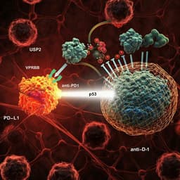
Targeting USP2 regulation of VPRBP-mediated degradation of p53 and PD-L1 for cancer therapy
J. Yi, O. Tavana, et al.

Medicine and Health
Comparing machine learning algorithms for predicting ICU admission and mortality in COVID-19
S. Subudhi, A. Verma, et al.


