logo
ResearchBunny Logo
Quantifying arousal and awareness in altered states of consciousness using interpretable deep learning

Medicine and Health

Quantifying arousal and awareness in altered states of consciousness using interpretable deep learning

M. Lee, L. R. D. Sanz, et al.

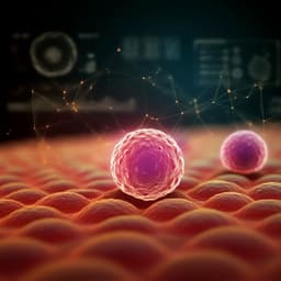
This groundbreaking research introduces the explainable consciousness indicator (ECI), utilizing deep learning to differentiate between arousal and awareness in various altered states of consciousness. By analyzing EEG responses in patients undergoing sleep, anesthesia, and severe brain injuries, the ECI reveals fascinating insights into states like ketamine-induced anesthesia, revealing the complexity of human consciousness. Discover the work of leading experts, including Minji Lee and Steven Laureys, in this innovative study.

00:00
00:00
~3 min • Beginner • English
Introduction
The study addresses the challenge of objectively quantifying consciousness by disentangling its two components: arousal (wakefulness) and awareness (subjective experience). Traditional clinical assessments conflate responsiveness with consciousness, despite evidence that subjective experiences can occur during unresponsive states such as certain sleep stages or anesthesia. Existing neurophysiological metrics like the perturbational complexity index (PCI) distinguish conscious from unconscious states but cannot separate arousal from awareness, and require many trials. Spectral EEG measures can reflect arousal but incompletely capture awareness. The authors propose an interpretable deep learning-based Explainable Consciousness Indicator (ECI) that outputs two probabilities representing arousal and awareness, aiming to classify and quantify these components across physiological (sleep/wake), pharmacological (propofol, xenon, ketamine anesthesia), and pathological (disorders of consciousness) conditions, including validation against PCI and extension to resting-state EEG.
Literature Review
Prior work established PCI from TMS-EEG as a validated measure to discriminate consciousness vs. unconsciousness with a cutoff near 0.31 but unable to distinguish REM sleep or ketamine anesthesia from wakefulness and requiring multiple trials. Resting-state EEG metrics like the spectral exponent correlate with PCI and can separate ketamine from propofol/xenon but have limitations distinguishing awareness across states and can overlap between conditions. REM vs. wakefulness discrimination using high-frequency spectral slopes reflects arousal more than awareness. Machine learning, especially CNNs, enables single-trial EEG classification but traditionally lacks interpretability; Layer-wise Relevance Propagation (LRP) provides explanations of model decisions. This background motivates a data-driven, interpretable approach leveraging spatiotemporal EEG features to separately classify arousal and awareness.
Methodology
Design: Developed a deep learning framework to compute a 2D Explainable Consciousness Indicator (ECI) comprising ECI_aro (arousal) and ECI_awa (awareness), each representing the averaged probability (0–1) of high arousal or high awareness for a session. The approach was applied to TMS-EEG and resting-state EEG and validated across sleep, anesthesia, and disorders of consciousness (DoC). Datasets: TMS-EEG: sleep (n=6 healthy; REM with dreams, NREM without dreams, and wake), anesthesia (n=16 healthy; ketamine n=6, propofol n=5, xenon n=5) with pre-anesthesia wake, DoC patients (UWS n=15, MCS n=15, MCS* n=4). Resting-state EEG: anesthesia (ketamine n=5, propofol n=5, xenon n=5) with pre-anesthesia wake; DoC (MCS n=15, UWS n=15, MCS* n=4). EEG acquisition: 60-channel TMS-compatible EEG (plus EOG), 1450 Hz sampling; TMS applied mainly over parietal/motor/premotor (sleep: parietal), with white noise masking and foam to reduce artifacts; resting-state EEG recorded at matching time points. Preprocessing: Downsample to 362.5 Hz; 0.5–45 Hz band-pass; segment TMS-EEG −400 to 1000 ms around TMS; ICA removal of ocular components; bad channel interpolation; trials exceeding ±100 μV rejected; average reference. Resting EEG segmented into 1 s epochs; first 200 ms used. Feature construction: Converted 1D multichannel EEG to 2D spatial meshes (10×11 grid) per time point; formed 3D inputs using spatiotemporal windows (TMS-EEG: 200–400 ms post-TMS yielding 10×11×72; resting EEG: first 200 ms) or spatio-spectral (five bands: delta, theta, alpha, beta, gamma; 10×11×5). Classifiers: Custom CNN with five convolutional layers, ReLU activations, max pooling, and final softmax for binary classification (low vs. high) separately for arousal and awareness. Training: Leave-one-participant-out (LOPO) cross-domain transfer learning: domains defined as sleep, anesthesia, DoC. Source domains selected based on cosine similarity; models trained on source domains excluding the target participant. For sleep/anesthesia targets, pooled similar domains; for DoC, trained with DoC plus anesthesia. Baselines included LDA and SVM (polynomial kernel). ECI computation: For each session, average the softmax probability of the high class across trials to obtain ECI_aro (x-axis) and ECI_awa (y-axis); threshold of 0.5 used to dichotomize. Validation: ROC analyses for sensitivity, specificity, AUC; correlations with PCI (sessions with ≥80 trials), and between TMS-derived ECI and resting-state ECI. Assessed performance vs. number of trials (1–80). Generalization to unseen patients via a hold-out split (DoC 75/25). Interpretability: Applied LRP to compute relevance scores over scalp regions (frontal, temporal, parietal) to identify spatial contributions; additional analyses removing regional electrodes to test impact on performance. Statistical analyses included Kruskal–Wallis tests, post hoc t-tests with Fisher’s LSD, and Pearson correlations.
Key Findings
- ECI separates arousal and awareness across conditions: NREM sleep and propofol/xenon anesthesia show low arousal and low awareness; REM sleep and ketamine anesthesia show low arousal with high awareness; wakefulness shows high arousal and high awareness; MCS patients show high arousal and high awareness; UWS patients show high arousal and low awareness. - ROC performance for TMS-EEG ECI: Sleep/wake: arousal AUC=1.0 (sensitivity=1.0, specificity=1.0); awareness AUC=0.995 (low awareness sensitivity=1.0, specificity=0.98; high awareness sensitivity=0.98, specificity=1.0). Anesthesia: AUC, sensitivity, specificity all 1.0 for both arousal and awareness. DoC: awareness AUC=1.0 (both high and low awareness); arousal ROC not applicable (both UWS and MCS considered high arousal). - Single-trial feasibility: In sleep/wake, using 1 trial achieved AUC=0.931, sensitivity=0.884, specificity=0.853; with ≥2 trials, all metrics exceeded 0.9. - Cross-domain single-trial classification accuracies (mean±SD) for TMS-EEG (LOPO transfer learning): Sleep target with Sleep source: arousal 87.79±2.50%, awareness 91.95±4.74%; Anesthesia target with Anesthesia source: arousal 79.01±10.61%, awareness 80.20±10.06%; DoC target with DoC+Anesthesia source: arousal 83.12±12.79%, awareness 78.78±12.98%. - Resting-state EEG classification accuracies: Anesthesia target with Anesthesia source: arousal 89.91±8.01%, awareness 90.14±6.96%; DoC target with DoC source: arousal 73.33±14.25%, awareness 88.62±8.83%; mixed-domain training improved specific components. - Strong correlation between ECI_awa and PCI across states: sleep r=0.872 (p<0.001), anesthesia r=0.885 (p<0.001), brain-injury patients r=0.770 (p<0.001). - Resting-state vs TMS-EEG ECI correlations: Anesthesia ECI_aro r=0.848 (p<0.001), ECI_awa r=0.938 (p<0.001); DoC ECI_aro r=0.534 (p=0.02), ECI_awa r=0.832 (p<0.001). - Generalization without retraining: Hold-out evaluation in DoC showed high correlation with LOPO-derived ECI (ECI_aro r=0.702, p=0.05; ECI_awa r=0.886, p<0.001) and correct categorization of MCS (high awareness) vs UWS (low awareness). - Interpretability: LRP revealed higher relevance over parietal regions than frontal or temporal for both arousal and awareness across sleep, anesthesia, and DoC, including non-parietal TMS targets. Removing parietal electrodes reduced ECI_awa AUC in DoC from 1.0 to 0.680 (frontal removal to 0.867), highlighting parietal importance. - MCS* patients (behaviorally UWS, metabolic MCS) were correctly predicted as high awareness by ECI in both TMS-EEG and resting-state EEG.
Discussion
The ECI framework addresses the core challenge of disentangling arousal and awareness by learning spatiotemporal EEG patterns that reflect these components across diverse altered states. It successfully classifies states that traditional indices like PCI cannot separate (e.g., REM and ketamine vs wakefulness) and does so with minimal data, enabling near single-trial inference. Strong correlations with PCI validate ECI_awa as a consciousness-related measure, despite methodological differences, supporting the reliability of deep learning-derived indicators. The parietal dominance in LRP relevance aligns with the posterior hot zone hypothesis, suggesting that EEG features over parietal cortex carry decisive information about levels of consciousness in both physiological and pathological contexts. Clinically, ECI’s accurate performance using resting-state EEG extends applicability beyond TMS-EEG, facilitating bedside assessments in anesthesia monitoring and DoC diagnosis. Transfer learning across related domains and leave-one-participant-out training demonstrate participant-independent generalization, important for translational use.
Conclusion
The study introduces an interpretable deep learning-based Explainable Consciousness Indicator that simultaneously quantifies arousal and awareness from EEG, validated across sleep, anesthesia, and disorders of consciousness. ECI achieves high accuracy, correlates strongly with PCI, distinguishes REM/ketamine states from wakefulness, and works with few trials and even without TMS using resting-state EEG. LRP-based interpretation consistently implicates parietal regions in decision-making, supporting posterior hot zone theories and suggesting streamlined electrode configurations for clinical deployment. Future directions include validating on larger cohorts, implementing real-time ECI computation, refining domain/model selection for deployment, and developing ECI into a functional index capable of capturing qualitative aspects of conscious content.
Limitations
- Modest sample sizes across conditions; broader validation on larger cohorts is needed, particularly for sleep experiments. - Real-time computation was not implemented; ECI feasibility with true online processing remains to be demonstrated. - ECI distinguishes levels of arousal and awareness but does not identify the specific physiological, pharmacological, or pathological etiology of a state; model selection requires knowledge of domain during deployment. - Potential contamination of TMS-EEG by residual auditory/somatosensory co-stimulation, though mitigated by noise masking and coil padding; cannot be fully excluded. - ECI is not a functional measure of consciousness content; further development is needed to assess functional aspects. - Many TMS targets were parietal, although analyses with non-parietal targets and resting EEG still indicated parietal relevance; residual bias cannot be fully ruled out.
Listen, Learn & Level Up
Over 10,000 hours of research content in 25+ fields, available in 12+ languages.
No more digging through PDFs, just hit play and absorb the world's latest research in your language, on your time.
listen to research audio papers with researchbunny