Medicine and HealthNature Communications
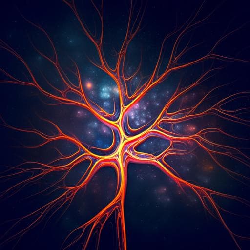
Physics-informed deep generative learning for quantitative assessment of the retina
E. E. Brown, A. A. Guy, et al.
Discover a groundbreaking algorithmic approach that revolutionizes the generation of realistic digital models of human retinal blood vessels. This innovative method, developed by Emmeline E. Brown and colleagues, surpasses traditional labeling performance through physics-informed generative adversarial networks, paving the way for enhanced early detection and monitoring of retinal diseases.
Related Publications
Explore these studies to deepen your understanding
Adjacent work that informs or extends this paper's methodology and findings.

Medicine and Health
Development and evaluation of deep learning algorithms for assessment of acute burns and the need for surgery
C. Boissin, L. Laflamme, et al.

Education
The role of teacher patience in the implementation of assessment for learning (AfL): Vignettes from a writing classroom
X. Zhang
Medicine and Health
A multimodal deep learning approach for the prediction of cognitive decline and its effectiveness in clinical trials for Alzheimer’s disease
C. Wang, H. Tachimori, et al.

Earth Sciences
Deep multi-task learning for early warnings of dust events implemented for the Middle East
R. Sarafian, D. Nissenbaum, et al.


