
Medicine and HealthCommunications Physics
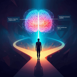
Personalized predictions and non-invasive imaging of human brain temperature
D. Sung, P. A. Kottke, et al.
This groundbreaking research conducted by Dongsuk Sung and colleagues unveils a biophysical model that predicts personalized brain temperature distributions using MRI data, laying the foundation for enhanced recovery after brain injuries. The study successfully validates this model against experimental measurements, promising significant implications for clinical applications in brain health.
Related Publications
Explore these studies to deepen your understanding
Adjacent work that informs or extends this paper's methodology and findings.
Psychology
Decision-Making with Predictions of Others’ Likely and Unlikely Choices in the Human Brain
N. Ma, N. Harasawa, et al.

Engineering and Technology
Ultra-high gradient connectomics and microstructure MRI scanner for imaging of human brain circuits across scales
G. Ramos-llordén, H. Lee, et al.

Medicine and Health
Non-invasive modulation of meningeal lymphatics ameliorates ageing and Alzheimer's disease-associated pathology and cognition in mice
M. Wang, C. Yan, et al.

Medicine and Health
Cardiovascular and autonomic dysfunction in long-COVID syndrome and the potential role of non-invasive therapeutic strategies on cardiovascular outcomes
G. Pérez-rubio, R. D. Rio, et al.


