
Biology
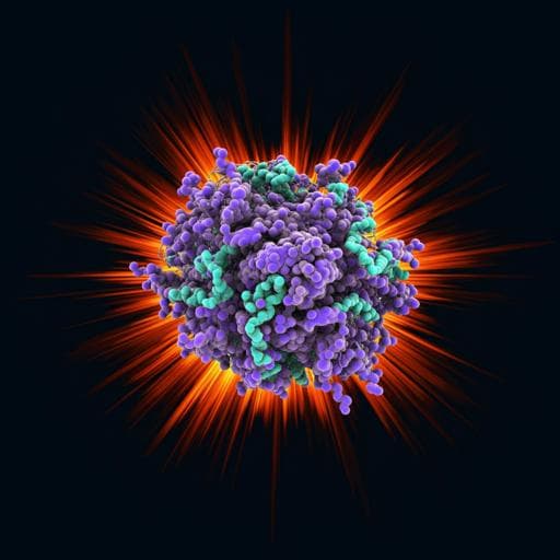
Observation of a single protein by ultrafast X-ray diffraction
T. Ekeberg, D. Assalaoua, et al.
Discover the groundbreaking achievement of capturing an X-ray diffraction pattern from a single protein, *Escherichia coli* GroEL, by a team of researchers led by Tomas Ekeberg and others. This innovation enables ultrafast, time-resolved studies on a femtosecond timescale—revolutionizing how we understand protein dynamics.
~3 min • Beginner • English
Introduction
X-ray free electron lasers (XFEL) have transformed the study of ultrafast phenomena at the atomic level, from transient room-temperature superconductivity to the fastest processes following water ionisation. This has also been the case in structural biology with the birth of serial femtosecond crystallography (SFX) and more recently the development of time-resolved SFX. Yet the requirement of crystals is limiting as demonstrated by the spectacular development in cryo-electron microscopy (cryo-EM). More importantly, the need to synchronise all unit cells in a crystal makes photo-activation the only feasible trigger for ultrashort timescales. It also prevents the observation of individual molecular behaviour, e.g., multiple conformations. Currently, cryo-EM is the method of choice for high-resolution single-molecule time-resolved studies, but it is limited to millisecond timescales due to the time it takes to freeze the sample and collect the data. By bypassing these limitations, femtosecond X-ray diffractive imaging (FXI) has the potential to observe single molecules with sub-picosecond time resolution, and due to the higher sample temperature, may allow sampling from a broader conformational landscape.
The chaperonin GroEL is an abundant molecular chaperone and, together with its cofactor GroES, is important in the folding of a large range of proteins. Escherichia coli GroEL is a 14-mer formed by two heptameric subunit rings, totaling ~800 kDa and arguably the most studied chaperonin. It was also one of the first large macromolecular complexes to be successfully measured by native mass spectrometry and nowadays often used as a benchmark to demonstrate the resolution of new systems. Its size and availability also made it an early target for single-particle cryo-EM studies. These characteristics along with the extensive body of available knowledge and distinctive shape, recognisable even at low resolution, make GroEL an ideal prototype system for single-particle X-ray diffraction.
Despite continuous progress in FXI, no single-particle studies have ever been measured, and studies involving GroEL have focused on larger complexes. It can, however, serve as the first interpretable X-ray diffraction signal from a protein complex, the chaperonin GroEL, and arguably it is lighter than the smallest biological sample previously reported, the ~9 MDa Toxoplasma busy stunt virus. With it we demonstrate the principle of diffraction before destruction. This opens the doors to ultrafast studies on single-protein molecules making use of the extraordinary brightness and time resolution of XFELs.
Literature Review
Methodology
The experiment was conducted at the Small Quantum Systems (SQS) instrument of the European XFEL (EuXFEL) in Schenefeld, Germany. GroEL protein particles were injected into the gas phase and intersected with femtosecond soft X-ray pulses from the EuXFEL at a photon energy of 1200 eV and an average pulse energy of 6.5 mJ.
Sample preparation and injection: Individual GroEL particles were transferred from solution to the gas phase using an electrospray setup. A charged liquid jet produced droplets of approximately 110 nm diameter in an inert CO2/N2 gas sheath. Droplets were then neutralised and focused by an aerodynamic lens, creating a thin particle beam. During transit most or all volatile buffer evaporated, yielding a stream of mostly dry particles delivered to the XFEL interaction region.
Detection and geometry: Diffraction was recorded on a pnCCD detector comprising two detection planes located 150 mm downstream of the interaction point. The setup’s numerical aperture imposed a resolution limit of about 4 nm. The detector’s energy resolution (~40 eV) enabled discrimination of elastic scattering at 1200 eV from fluorescence photons at 277 eV (C Kα1), 392 eV (N Kα1), and 525 eV (O Kα1) for pixels receiving at most one photon.
Background characterization: Background arose from the injection gas (both fluorescence and elastic scatter) and the beamline. Control measurements with the injector off and with gas on but no sample quantified contributions: the injection gas contributed on average ~17,600 photons per diffraction pattern, whereas the beamline contributed ~86 photons per pattern on average.
Data acquisition constraints: EuXFEL delivers pulse trains at 10 Hz with MHz repetition within trains. The CCD-based detector allowed only a single image readout per train, limiting acquisition. From 84,000 total readouts, 816 patterns passed initial hit detection; many were background fluctuations. Patterns exceeding 19,000 photons (172 patterns) predominantly contained true sample hits.
Classification and size estimation: Diffraction patterns were compared against sphere models of varying sizes; a histogram of events with >19,000 photons peaked at ~15 nm diameter, consistent with GroEL dimensions (~14 nm), with a secondary small peak below 5 nm attributed to strong background shots.
Single-pattern analysis and template matching: A representative high-signal pattern exhibiting clear deviation from circular symmetry (consistent with GroEL’s barrel shape) was analysed. To address unknown molecular orientation and pattern centre, a template-matching approach was used: diffraction from a GroEL structural model was simulated across orientations sampling full 3D reciprocal space in ~7° steps, and translated across possible pattern centres. In total, 4.3 million simulated and translated patterns were compared against the experimental pattern. Background handling involved using average background patterns from runs with injection gas but no sample to separate signal from background.
Model comparisons: Additional models augmented the GroEL structure with water-like density to test the influence of residual solvent and internal cavity filling; models varied from ~13% to ~69% added mass relative to protein, with all but one filling GroEL’s central cavity.
Key Findings
- First observation of an X-ray diffraction pattern from a single protein molecule (Escherichia coli GroEL), demonstrating diffraction-before-destruction at the single-protein level and constituting the smallest biological sample imaged by X-rays (~14–15 nm diameter).
- Experimental conditions: 1200 eV photon energy, ~6.5 mJ average pulse energy, pnCCD detection with two planes 150 mm downstream, ~4 nm resolution limit due to numerical aperture.
- Background quantification: injection gas contributed ~17,600 photons per pattern on average; beamline contributed ~86 photons per pattern; detector energy resolution (~40 eV) allowed separation of fluorescence from elastic scattering for single-photon pixels.
- Data yield: Of 84,000 readouts, 816 triggered as hits; 172 patterns contained >19,000 photons and were predominantly true hits.
- Size estimation from diffraction indicated a strong peak at ~15 nm for patterns with >19,000 photons, matching GroEL’s expected size; a smaller peak <5 nm corresponded to strong background shots.
- A selected high-signal single-particle diffraction pattern showed anisotropic first fringe consistent with GroEL’s barrel geometry; template matching across ~4.3 million simulated orientations/translations enabled approximate orientation determination and supported assignment to GroEL.
- Comparisons with models including added water-like density explored the effect of residual solvent and cavity filling on the diffraction signal.
Discussion
The study addresses the long-standing goal of obtaining single-molecule structural information without crystals by exploiting ultrashort, intense XFEL pulses. By recording and identifying a diffraction pattern from a single GroEL protein, the work validates that sufficient coherent elastic scattering can be captured before radiation damage destroys the sample, thus demonstrating diffraction before destruction at the protein scale. The observed deviation from circular symmetry in the experimental pattern and successful template matching to a GroEL model establish both the molecular origin of the signal and the feasibility of approximate orientation recovery from single-shot data.
These results are significant for structural biology and ultrafast science: they remove the crystal requirement, enabling observations of individual conformational states and the possibility to follow dynamics at femtosecond timescales at or near room temperature. Quantification of background contributions and a practical strategy for background discrimination and hit classification provide a roadmap for future experiments. While the current resolution and data rate limit detailed structure determination, the demonstrated signal levels, orientation sensitivity, and size consistency make single-molecule femtosecond X-ray diffractive imaging a realistic path toward time-resolved studies of macromolecular dynamics.
Conclusion
This work reports the first X-ray diffraction pattern from a single protein (GroEL), establishing that diffraction-before-destruction extends to single macromolecules and that approximate orientation can be extracted from a single shot. The experiment quantifies key practicalities—background levels, hit rates, and instrument constraints—while demonstrating data-analysis strategies (template matching across millions of orientations/translations and background handling) that enable interpretation. These achievements open the way to ultrafast, single-molecule, time-resolved investigations on femtosecond timescales, potentially accessing a wider conformational landscape of proteins at near-physiological temperatures. Future work can focus on improving data rates (e.g., faster detectors), reducing background (optimized injection and beamline cleaning), extending to other proteins and states (including GroEL-GroES complexes), and pushing toward higher resolution and full 3D reconstructions from single-shot ensembles.
Limitations
- Detector readout limited to one image per XFEL pulse train (CCD technology), severely constraining data collection rate and leading to low overall hit counts (816/84,000 initial detections; 172 strong-hit patterns >19,000 photons).
- High background from injection gas (average ~17,600 photons per pattern) necessitated stringent discrimination; residual background fluctuations caused false hits near the background peak (~17,000 photons).
- Instrumental resolution limited to ~4 nm by the detector’s numerical aperture, precluding atomic-resolution structural information from the present dataset.
- Single-shot analysis required handling unknown pattern centre and molecular orientation; reliance on extensive template matching (4.3 million simulations) introduces model dependence and computational demands.
- Data presented focus on a single favourable diffraction pattern for detailed analysis; broader statistical validation and reconstructions from multiple orientations were beyond the scope of the reported acquisition.
- Background separation using energy discrimination is effective only for pixels receiving at most one photon, limiting its applicability at higher local intensities.
Related Publications
Explore these studies to deepen your understanding of the subject.