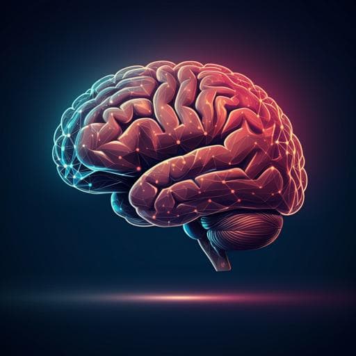
Neural foundation of the diathesis-stress model: longitudinal gray matter volume changes in response to stressful life events in major depressive disorder and healthy controls
F. Thomas-odenthal, K. Ringwald, et al.
Explore these studies to deepen your understanding
Adjacent work that informs or extends this paper's methodology and findings.
Acute changes in antioxidants and oxidative stress to vigorous arm exercise: an intervention trial in persons with spinal cord injury and healthy controls
M. F. Wouda, H. B. Steihaugell, et al.
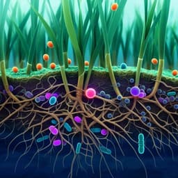
The response of wheat and its microbiome to contemporary and historical water stress in a field experiment
H. Azarbad, L. D. Bainard, et al.

Obesity and adverse childhood experiences in relation to stress during the COVID-19 pandemic: an analysis of the Canadian Longitudinal Study on Aging
V. D. Rubies, A. Gonzalez, et al.

Insights to the neural response to food cues in class III compared with class I and II obese adults using a sample of endometrial cancer survivors seeking weight loss
N. L. Nock, H. Jiang, et al.


