Medicine and Health
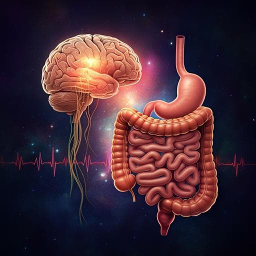
Multifunctional microelectronic fibers enable wireless modulation of gut and brain neural circuits
A. Sahasrabudhe, L. E. Rupprecht, et al.
Discover groundbreaking multifunctional neural interfaces merging advanced polymer-based fibers with microelectronics to explore the depths of brain and gut neurophysiology. This revolutionary research by Atharva Sahasrabudhe and colleagues paves the way for new insights into interoception and behavior modulation.
~3 min • Beginner • English
Introduction
The study addresses the challenge of interrogating bidirectional brain–viscera communication, particularly gut–brain pathways, using implantable devices that can safely and chronically operate in anatomically and mechanically disparate organs. Traditional lithography-based neural interfaces require separate fabrication and manual assembly of different modalities, limiting rapid customization and scalability. The authors propose thermally drawn, polymer-based microelectronic fibers that monolithically integrate optical, electrical, thermal sensing, and microfluidic functions with tunable mechanics suitable for both deep brain and gastrointestinal applications. The purpose is to enable wireless, programmable neuromodulation and physiological recording in freely behaving animals to uncover mechanisms by which gut signals influence central neural circuits and behavior.
Literature Review
The paper notes limitations of traditional thin-film, lithography-based neural interfaces that require resource-intensive fabrication and manual multimodal assembly, hindering scalability and customization for diverse anatomical targets. Prior works on flexible, multimodal, and wireless optoelectronics for neuromodulation are cited, as well as thermally drawn multimaterial fiber electronics. The need remains for monolithic, scalable, multifunctional bioelectronic interfaces with long-term biocompatibility and design flexibility across organs such as the brain and gut.
Methodology
Device fabrication and architecture: Multifunctional fibers were produced via thermal drawing from macroscopic preforms.
- Brain fibers: Polycarbonate (PC) multilayer preform (Tg = 150 °C, E = 1.8–3.2 GPa) with channels for interconnects, electrodes, and microfluidics was thermally drawn while simultaneously feeding Ag–Cu interconnect microwires (40 µm) and tungsten electrode microwires (25 µm). Post-draw, surface-mounted blue (470 nm) and green (527 nm) InGaxN µLEDs (270 × 210 × 50 µm³) were bonded on the fiber, and devices were parylene-C coated. Fibers integrated: (1) surface-localized µLEDs for optogenetics; (2) tungsten microelectrodes for electrophysiology; (3) thermal sensing using µLED I–V thermometry; (4) microfluidic channel for gene/chemical delivery.
- Gut fibers: Soft, compliant fibers using SEBS elastomer cladding (Tg = 140 °C, E = 3–5 MPa), with carbon-doped polyethylene (CPE) conductors and a thin PC layer for structural integrity. Convergence drawing incorporated Ag–Cu interconnects. Final devices (~8.5 cm) hosted six surface-mounted µLEDs (three blue, three green) arranged along distal 2 cm, and a microfluidic outlet ~0.8–1 mm proximal to the first µLED pair. Devices were encapsulated in parylene-C and medical-grade silicone.
Characterization: Measured electrode impedance spectra in PBS, µLED optical output versus power and bending, FEM for optical intensity/penetration and thermal rise, microfluidic return rates, bending stiffness (single-cantilever), and mechanical FEM for micromotion and stress/strain. Gut fiber robustness was tested via cyclic buckling and bending; strain in copper interconnects under tight bending radii was simulated.
Wireless module (NeuroStack): A 15.5-mm, 1.1-g modular BLE platform (MDBT42V with Nordic nRF52832) enabling programmable optical stimulation (frequency, duty cycle, pulse shape/intensity) across two channels and real-time temperature recording (default 200 Hz sampling). An optional intensity module (DAC MAX5510) provided analog intensity control and pulse shaping. Power from rechargeable batteries; initial brain devices operated up to ~1 h on 11 mAh battery, initial gut devices ~30 min; a modified version supported up to ~2 h with a 45 mAh battery.
In vivo procedures:
- Brain: One-step stereotaxic implantation of VTA-targeting brain fibers in DAT::Cre mice, with AAV5 Ef1α::DIO-ChR2-mCherry or control DIO-mCherry delivered via fiber microfluidics during implantation. Longitudinal opto-electrophysiology recorded spontaneous and optically evoked activity; thermal sensors recorded intracranial temperature. Biocompatibility assessed by Iba1 and GFAP immunohistochemistry.
- Gut: Intraluminal implantation of soft gut fibers in duodenum or ileum via purse-string sutures; distal fiber threaded via pylorus (duodenum) or distal ileum (near cecum). Wireless intraluminal optogenetics targeted Cck+ neuropod cells (duodenum), Pyy+ cells (ileum), and upper-gut Phox2b+ vagal afferents; microfluidic nutrient delivery (e.g., sucrose) was performed. Vagal nerve cuff recordings quantified afferent responses.
- Behavioral assays: Real-time place preference (RTPP) with wireless programmable photostimulation in VTA (DAT::Cre) and gut (Phox2b::ChR2). Feeding assays: chow intake after duodenal Cck::ChR2 stimulation, Ensure solution intake after ileal Pyy::ChR2 stimulation. Concurrent VTA electrophysiology during intraduodenal sucrose vs saline delivery assessed DA neuron responses.
Data acquisition and analysis: Electrophysiology sampled at 50 kHz, filtered 0.3–5 kHz; spikes detected at 5 s.d. threshold; PCA and GMM clustering for single units; L-ratio and isolation distance for cluster quality. Wireless temperature recorded via digitized sensor currents; calibration using hotplate/thermocouple. Statistical analyses included ANOVA, t-tests, Kruskal–Wallis, and Wilcoxon tests as appropriate.
Key Findings
Device performance and mechanics:
- Brain fibers: Tungsten microelectrodes exhibited low impedance ZI = 46.3 ± 6 kΩ at 1 kHz, stable over 7 weeks in PBS; no leakage through cladding. Blue µLED output tunable from 0.6 to 70 mW mm−2; robust to bending up to 90° with stable output for ≥7 weeks in PBS. FEM showed 30 mW mm−2 illuminates ~0.75 mm³ near surface. Thermal sensor detected negligible ΔT = 0.085 °C from adjacent blue µLED (≈30 mW mm−2, 40 Hz). Microfluidics showed 80–100% return rate at 20–100 nl s−1. Bending stiffness 25–33 N m−1, markedly lower than silica (132 N m−1) and steel (792 N m−1); FEM predicted 2–4 orders lower relative micromotion than silica/steel.
- Gut fibers: Optical penetration depth 0.15–1 mm; illumination volume 0.9–8.8 mm³ at 20–100 mW mm−2; negligible heating; µLED spacing (~1 cm) avoided cooperative heating. Robust to cyclic buckling (1–5 mm, 10 cycles) and bending (90°/180°; minimal output change at 2–10 cm radii). Bending stiffness 2–5 N m−1 (vs PC 70–75 N m−1; silica 132 N m−1). In vivo, rigid silica punctured mucosa, while soft fibers implanted without damage. Interconnect strain remained below elastic limit (~0.3%) for radii >0.5 cm. Microfluidic return rates 60–90% at 1–5 µl s−1 in straight and bent states.
Wireless platform (NeuroStack): Enabled real-time control of frequency (10–40 Hz), duty cycle (20–80%), pulse shape (rise/fall 5–15 ms), and intensity; BLE link with on-board battery; wireless temperature sensing linear with steady-state temperatures (29–40 °C) and dynamics matched a thermocouple.
In vivo brain validation (VTA, DAT::Cre):
- AAV5 ChR2 expression achieved via one-step microfluidic delivery. Emergence of optically evoked activity tracked opsin expression over weeks; multiunit activity recorded ≥2 months; single units recorded up to 4 weeks; electrodes functional up to 6 months. Optical artifact mitigation via pulse shaping validated.
- Wireless intracranial temperature showed no significant heating during photostimulation; thermal sensors detected anesthesia-induced brain hypothermia (ketamine-xylazine): dose-dependent drops at 30 and 60 mg kg−1 ketamine.
- Behavioral RTPP with wireless photostimulation: ChR2-mCherry mice showed significant preference for stimulated chamber; mCherry controls did not.
• On vs OFF: ChR2-mCherry P = 4.16×10⁻⁹, t = −9.03, d.f. = 7; mCherry P = 0.183, t = −1.50, d.f. = 6.
• Phasic vs tonic: ChR2-mCherry P = 2.02×10⁻⁷, t = −9.65, d.f. = 5; mCherry P = 0.403, t = −0.88, d.f. = 7.
• Blue vs green: ChR2-mCherry P = 3.58×10⁻⁵, t = −9.24, d.f. = 7; mCherry P = 0.55, r = −0.64, d.f. = 5.
No differences in locomotion vs naïve mice; µLEDs remained functional ≥9 months.
In vivo gut validation and gut–brain:
- Vagal electrophysiology: In wild-type mice, intraduodenal sucrose (300 mM, 0.2 ml, 3.3 µl s−1) increased vagal firing vs baseline (Kruskal–Wallis, P = 0.0304). In Cck::ChR2 mice, duodenal blue µLED stimulation (40 Hz, 10 ms) increased vagal firing; green did not; controls (no ChR2) showed no change. Peak response comparisons: baseline vs blue P = 0.0367 (n = 5); baseline vs green P = 0.1113 (n = 4); blue vs green P = 0.0200.
- Feeding behavior:
• Duodenum (Cck::ChR2): Blue-light (20 Hz, 10 ms; 0.5 s ON/1 s OFF) significantly suppressed chow intake vs green within-subjects over 3 h (n = 4): effects of time P = 0.0003, stimulation P < 0.0001, time×stimulation P = 0.0086; post hoc paired t-tests: 1 h P = 0.0161; 2 h P = 0.0376; 3 h P = 0.0044. Controls lacking ChR2: time P = 0.0020; stimulation P = 0.4975; interaction P = 0.8906.
• Ileum (Pyy::ChR2): Blue-light suppressed Ensure (30%) intake vs green (n = 4): time P < 0.0001, stimulation P < 0.0001, interaction P < 0.0001; cumulative intake paired t-test P = 0.0381. Controls (n = 4): time P < 0.0001; stimulation P = 0.0160; interaction P = 0.5796; cumulative intake P = 0.4639.
- Brain recording during gut nutrient delivery: In wild-type mice with VTA brain fibers, intraduodenal sucrose (0.6 M, 0.5 ml over 10 min) increased VTA putative DA neuron firing vs saline. Wilcoxon signed-rank: saline P = 0.8595 (n = 20 neurons), sucrose P = 6.71387×10⁻¹² (n = 18 neurons). DA identity supported by quinpirole (D2 agonist) sensitivity.
- Vagal afferent optogenetics (Phox2b::ChR2): Duodenal blue-light stimulation induced RTPP (paired t-test P = 0.014, t = 4.19, d.f. = 4); genetic controls NS (P = 0.110, t = −2.25, d.f. = 3).
Biocompatibility: Brain fibers evoked lower glial response (Iba1, GFAP) than comparable silica fibers at week 2; gut fibers did not alter villus/crypt morphology and did not obstruct food/water intake or locomotion.
Discussion
The work demonstrates that thermally drawn, polymer-based microelectronic fibers can monolithically integrate multimodal functions with tailored mechanics to address both rigid CNS and soft gastrointestinal environments. When paired with a modular wireless controller (NeuroStack), the platform enables programmable optogenetics and physiological recording in freely behaving mice. In the brain, targeted delivery of viral constructs, stable long-term electrophysiology, real-time temperature monitoring, and wireless optogenetic modulation of VTA DA neurons yielded robust reward-related behaviors, validating functionality and safety. In the gut, soft fibers enabled intraluminal, laterally directed light and nutrient delivery to sparsely distributed epithelial neuropod cells and to vagal afferents, eliciting cell-type-specific changes in vagal activity and measurable alterations in feeding behavior. Further, concurrent recordings showed that intestinal nutrient sensing can modulate VTA DA neuron firing, and optogenetic activation of vagal afferents from the lumen is sufficient to drive reward, directly linking gut sensory pathways to central motivational circuits. These findings address the lack of biointegrated tools for multi-organ neuromodulation and open avenues for dissecting interoceptive circuits relevant to metabolism, affect, and cognition.
Conclusion
This study introduces a scalable, monolithic, microelectronic fiber platform with tunable mechanics for wireless, multimodal neuromodulation and recording in both brain and gut. The fibers integrate optogenetics, electrophysiology, thermometry, and microfluidics, and are controlled by a lightweight BLE module enabling real-time programmability. In vivo, the system modulated VTA DA circuits to induce reward, controlled gut epithelial neuropod cells to alter feeding, and demonstrated that luminal activation of vagal afferents is rewarding. These capabilities provide a powerful toolkit for probing and manipulating gut–brain communication. Future work could extend functionalities (additional sensors/actuators), increase battery life and miniaturization for longer studies, adapt the platform to other organs and larger animals, and develop closed-loop paradigms coupling local sensing to adaptive stimulation for therapeutic applications.
Limitations
The paper does not provide a dedicated limitations section. Noted practical constraints include: (1) initial wireless gut device battery life (~30 min) necessitating hardware modification for longer assays (~2 h), with further extensions requiring larger-capacity batteries; (2) potential opto-electrophysiology artifacts on recording electrodes, mitigated via pulse shaping; (3) demonstrations are in mice and focused on selected brain (VTA) and gut regions (duodenum, ileum), with broader applicability to other circuits left for future studies.
Related Publications
Explore these studies to deepen your understanding of the subject.

